


In vitro incorporation of nonnatural amino acids into protein using tRNACys-derived opal, ochre, and amber suppressor tRNAs
- 1Department of Biochemistry and Molecular Biology, Oregon Health and Sciences University, Portland, Oregon 97231, USA
- 2Department of Molecular and Cellular Medicine, Texas A&M Health Science Center, College Station, Texas 77843-1114, USA
- 3Department of Chemistry, Texas A&M University, College Station, Texas 77843, USA
- 4Department of Biochemistry and Biophysics, Texas A&M University, College Station, Texas 77843, USA
Abstract
Amber suppressor tRNAs are widely used to incorporate nonnatural amino acids into proteins to serve as probes of structure, environment, and function. The utility of this approach would be greatly enhanced if multiple probes could be simultaneously incorporated at different locations in the same protein without other modifications. Toward this end, we have developed amber, opal, and ochre suppressor tRNAs derived from Escherichia coli, and yeast tRNACys that incorporate a chemically modified cysteine residue with high selectivity at the cognate UAG, UGA, and UAA stop codons in an in vitro translation system. These synthetic tRNAs were aminoacylated in vitro, and the labile aminoacyl bond was stabilized by covalently attaching a fluorescent dye to the cysteine sulfhydryl group. Readthrough efficiency (amber > opal > ochre) was substantially improved by eRF1/eRF3 inhibition with an RNA aptamer, thus overcoming an intrinsic hierarchy in stop codon selection that limits UGA and UAA termination suppression in higher eukaryotic translation systems. This approach now allows concurrent incorporation of two different modified amino acids at amber and opal codons with a combined apparent readthrough efficiency of up to 25% when compared with the parent protein lacking a stop codon. As such, it significantly expands the possibilities for incorporating nonnative amino acids for protein structure/function studies.
Keywords
- suppressor tRNA
- cysteine modification
- cell-free translation
- nonnatural amino acids
- tRNA aminoacylation
- translation termination
INTRODUCTION
Site-specific incorporation of nonnatural amino acids is a powerful tool used to manipulate proteins for structural and functional studies, or to create proteins with new properties (Wang and Schultz 2004). This is usually accomplished by one of two methods: (1) labeling presynthesized proteins with probes at reactive side chains such as cysteine or lysine residues or (2) incorporating probes into nascent proteins as they are being synthesized in the presence of a modified aminoacyl-tRNA (aa-tRNA) (Hendrickson et al. 2004; Wang et al. 2009). The first method generally requires extensive mutagenesis to remove redundant labeling sites and/or purification to eliminate labeled contaminants. In contrast, cotranslational incorporation using modified aa-tRNAs and an mRNA containing a unique cognate codon allows diverse probes to be positioned at virtually any site with high specificity and minimal perturbation of the protein sequence. The second approach most commonly utilizes an amber suppressor aa-tRNA that recognizes a unique nonsense (UAG) codon (Bain et al. 1989; Noren et al. 1989). However, a variety of synthetic and engineered aa-tRNAs have also been developed for this purpose, including those that recognize four-base codons (Hohsaka et al. 1996; Rodriguez et al. 2006).
To enable cotranslational incorporation, the nonnative amino acid must be attached to tRNA with high efficiency, either by using an engineered orthogonal aa-tRNA synthetase (aaRS) that recognizes both the modified amino acid and tRNA (Wang et al. 2009), or by chemical coupling to presynthesized tRNAs. While expression of an aaRS–tRNA pair enables probe incorporation in intact cells, generating suitable aaRS enzymes is labor intensive for higher eukaryotes and often limited to specific probe structures (Liu et al. 2007b; Chen et al. 2009). aa-tRNAs containing a modified amino acid can also be generated in vitro by chemical acylation of a probe to a synthetic dinucleotide and subsequent ligation to the 3′-end of a truncated tRNA (Heckler et al. 1984; Robertson et al. 2002; Ninomiya et al. 2003). Alternatively, coupling probes to an enzymatically aminoacylated tRNA is technically straightforward, and requires only that the tRNA is recognized by an aaRS and that the amino acid contains a chemically reactive side chain, such as a free amine (lysine) or a sulfhydryl group (cysteine) (Johnson et al. 1976; Flanagan et al. 2003). Enzymatic aminoacylation in vitro also provides the opportunity to create radiolabeled aa-tRNAs that allow accurate quantification of probe incorporation (Johnson et al. 1976).
The above approaches have enabled incorporation of photoactive cross-linkers (Liu et al. 2007b); azides/alkynes for “click”-chemistry (Liu et al. 2007b); photocaged residues (Chen et al. 2009); spin labels (Shafer et al. 2004); and fluorescent probes to measure binding affinities (Flanagan et al. 2003), protein structure (Woolhead et al. 2004), and environment (Crowley et al. 1993; Alder et al. 2008) in higher eukaryotic expression systems. Unfortunately, concomitant incorporation of two different nonnative amino acids at defined locations in the same protein has proven much more difficult. To date, this has been achieved using an amber suppressor tRNA in combination with a lysyl tRNA (Woolhead et al. 2004), a four-base anticodon tRNA and a suppressor tRNA (Rodriguez et al. 2006), and two different four-base anticodon tRNAs (Kajihara et al. 2006). However, each of these approaches requires removal of alternative incorporation sites, and the added modified aa-tRNAs must compete with endogenous aa-tRNAs.
Opal and/or ochre suppressor tRNAs are attractive candidates for probe incorporation because they require only the introduction of a unique stop codon into the coding sequence. Like amber suppressors, opal and ochre tRNAs are usually derived from a native scaffold in which the anticodon and/or adjacent bases have been mutated to pair with a UGA or UAA codon, respectively (Köhrer et al. 2004). tRNAPhe-derived opal and ochre tRNAs have been used to incorporate nitrophenylalanine in rabbit reticulocyte lysate (RRL) (Taira et al. 2005), and opal suppressors derived from tRNAGln or tRNATrp have been used to incorporate 5-hydroxytryptophan (Zhang et al. 2004) and 5-F-tryptophan (Rodriguez et al. 2007), respectively, in mammalian cells. Ochre suppressors derived from Escherichia coli supF tRNA and the suppressors of all three nonsense codons derived from E. coli tRNAGln have also been used to incorporate tyrosine (Köhrer et al. 2003) and glutamine (Köhrer et al. 2004) residues in mammalian cells, respectively. However, such efforts have been limited by the ability of the aaRS to recognize suppressor tRNAs and/or modified amino acids, and by low readthrough levels at their cognate codons. To date, concurrent suppression of two sequential nonsense codons has been disappointingly low (<2.5% readthrough) and has been achieved only using natural amino acids (Köhrer et al. 2004).
Here we describe a novel and generally applicable technique for the synthesis, aminoacylation, and modification of tRNACys derivatives that selectively recognize amber, opal, and ochre codons. These tRNAs enable readthrough at each of their cognate stop codons and allow sequential incorporation of two nonnatural amino acids in a eukaryotic translation system with an apparent efficiency up to 10-fold higher than previously reported. Our results establish a versatile means to incorporate multiple nonnatural amino acids at defined sites within a single protein.
RESULTS
Increased aminoacylation and stability of synthetic Cys-tRNAs
Because the sulfhydryl moiety of cysteine can be chemically modified with a wide variety of commercial compounds, tRNACys is a useful tool for cotranslationally incorporating unique probes at endogenous and introduced UGC codons (Alder et al. 2008). Cys-tRNAs from yeast and plants have also been used to incorporate Cys residues at opal (UGA) codons in vitro and in Xenopus oocytes (Vacher et al. 1984; Urban and Beier 1994). To establish tRNACys as a viable platform for developing suppressor tRNAs we first characterized E. coli, yeast, and human tRNACys (Lipman and Hou 1998) for their aminoacylation efficiency, aa-tRNA stability, and ability to incorporate a modified cysteine residue in two eukaryotic cell-free translation systems, wheat germ (WG) extract, and RRL. The tRNAs were synthesized by in vitro transcription and aminoacylated with [14C]Cys using an S-100 E. coli cytosolic extract. Although initial charging efficiencies were modest, aminoacylation was improved by the addition of purified recombinant E. coli and/or human cysteine aaRS (Table 1).
Aminoacylation of tRNAs
Resulting [14C]Cys-tRNAs were then purified and added to cell-free translation reactions programmed with a truncated mRNA transcript encoding a model protein [AQP4-P(98)] (Fig. 1) with a unique cysteine codon at residue 44 (Shi et al. 1995). Hot trichloroacetic acid (TCA) precipitation of translation products revealed poor incorporation of [14C]Cys when compared with incorporation of [14C]Lys at an equivalent amber (UAG) codon using a control amber suppressor tRNA, [14C]Lys-tRNALysamb (Fig. 2A). One reason for this was that Cys-tRNAs underwent rapid deacylation upon addition to the translation reaction. Notably, >50% of [14C]Cys-tRNACys was deacylated within 5 min in RRL (Fig. 2B) and 15 min in WG extract (Fig. 2C), whereas ∼50% of [14C]Lys-tRNALysamb remained intact after 1 h (Fig. 2B,C; Table 2). Some deacylation was also observed under equivalent buffer conditions (Fig. 2D). Thus, synthetic Cys-tRNAs are both less chemically stable than their Lys counterparts, and more susceptible to enzymatic breakdown, presumably by aaRS trans-editing activity in the WG extract and RRL (Jakubowski and Goldman 1992; Ahel et al. 2003; Ibba and Söll 2004).
Apparent half-lives of Cys-tRNAs in the RRL and WG extract cell-free translation systems
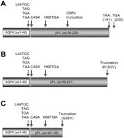
AQP4-P construct and mutations. (A) Cartoon of the fusion protein, AQP4-P, which contains the first 46 amino acids of human AQP4, a two-residue Val-Thr linker, and amino acids 88–229 of bovine prolactin (Shi et al. 1995). Codon 44 (Leu44) was mutated to an amber (TAG), opal (TGA), ochre (TAA) stop codon, or to Cys (TGC) as indicated. In the L44C mutant the endogenous Cys at residue 49 was also converted to Ala (C49A). For dual probe incorporation, amber and opal stop codons were simultaneously placed at residues Leu44 and His68, respectively. Where indicated, mRNA was truncated at codon 183 (B) or codon 98 (C) to generate the constructs AQP4-P(183) and AQP4-P(98), respectively. Full-length plasmids encode TAA at codon 191 and TGA at codon 200. Residues are numbered sequentially from Met1.
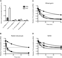
Incorporation and stability of Cys-tRNAs in RRL and WG extract translation systems. (A) The indicated aa-tRNAs were added to RRL (gray bars) or WG extract (white bars) translation reactions programmed with AQP4-P(98) cDNA containing a cognate cysteine (UGC) or an amber (UAG) codon at residue 44. [14C]Cys and [14C]Lys incorporation into protein was measured by liquid scintillation counting of hot acid precipitable counts. Results show mean ± SEM (n ≥ 3). (B–D) Deaminoacylation of Lys-tRNALysamb(•), yeast (▲), E. coli (■), or human (▼) Cys-tRNACys was determined by incubating aa-tRNAs in a mock translation reaction containing RRL (B), WG extract (C), or equivalent buffer (D). At the times indicated, aa-tRNA was precipitated in cold TCA and analyzed by scintillation counting. Counts obtained at t = 0 were used as a reference.
Cysteine modification enhances aa-tRNA stability and incorporation efficiency
To determine whether the cysteine side chain contributed to the poor stability of the aminoacyl-tRNA bond (Strickland and Jacobson 1972), we covalently attached a fluorescent moiety, 7-nitrobenz-2-oxa-1,3-diazole (NBD) to the sulfhydryl and the ɛ-amino group of [14C]Cys-tRNAs and [14C]Lys-tRNALysamb using iodoacetamide and succinimide ester derivatives, respectively. Modified tRNAs were then purified as described in Materials and Methods, which resulted in a highly enriched aa-tRNA population (specific activity = ∼1200 pm aa/A260U) that were efficiently (>90%) labeled on the respective Lys and Cys moieties. Notably, NBD attachment markedly improved stability of the Cys-tRNAs (Fig. 3C,D; Table 2), and correspondingly improved [14C]Cys incorporation efficiency approximately fivefold in WG extract (cf. Fig. 3A and Fig. 2A). In RRL, however, with the exception of yeast NBD-[14C]Cys-tRNACys, cysteine modification did not substantially change the rate of deacylation or [14C]Cys incorporation (Fig. 3A,B; Table 2). Thus, Cys-tRNA deacylation activity in RRL is also able to recognize and remove the NBD-modified Cys residue.
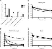
Incorporation and stability of NBD-Cys-tRNAs in RRL and WG extract translation systems. (A) The aa-tRNAs in Fig. 2A were modified with NBD as described in Materials and Methods and added to RRL (gray bars) or WG extract (white bars) translation reactions programmed with AQP4-P(98) cDNA containing a cognate cysteine (UGC) or amber (UAG) codon at residue 44. [14C]Cys and [14C]Lys incorporation into protein was measured by liquid scintillation counting of hot acid precipitable counts as described in Materials and Methods. Results show mean ± SEM (n ≥ 3). (B–D) Deaminoacylation of ɛNBD-Lys-tRNALysamb(•), yeast (▲), E. coli (■), or human (▼) NBD-Cys-tRNACys was determined as described in Fig. 2.
Incorporation of NBD-labeled Cys using Cys-derived suppressor tRNAs
We next mutated the GCA anticodon in yeast and E. coli tRNACys to CUA, UCA, or UUA to generate tRNACysamb, tRNACysopl, and tRNACysoch, respectively. While this nearly abolished aminoacylation by S-100 extract alone (Table 1), the addition of purified recombinant human Cys-aaRS restored aminoacylation of yeast-derived tRNAs, and the addition of either E. coli or human Cys-aaRS restored aminoacylation of E. coli-derived tRNAs (Table 1).
The stability of Cys-tRNACysamb, Cys-tRNACysopl, and Cys- tRNACysoch was similar to their WT counterparts (Table 2). Nonetheless, despite rapid deacylation, yeast Cys-tRNACysamb suppressed termination at the UAG codon with ∼25% efficiency of Lys-tRNALysamb in the WG extract, and the more stable derivative, NBD-Cys-tRNACysamb, suppressed termination with 70% efficiency compared with ɛNBD-Lys-tRNALysamb (Fig. 4A). NBD-Cys-tRNACysopl and NBD-Cys-tRNACysoch exhibited similar stability to NBD-Cys-tRNACysamb and also suppressed translation termination, although the extent of readthrough and probe incorporation was significantly less than at the amber codons (Fig. 4A; Table 2).
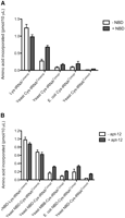
Incorporation of nonnatural amino acids by tRNACys-derived amber, opal, and ochre tRNAs. (A) Translation of AQP4-P(98) mRNA containing an amber, opal, or ochre codon at residue 44 was carried out in WG extract in the presence of cognate aa-tRNAs (white bars) or NBD-modified aa-tRNAs (gray bars). Incorporation of [14C]Lys or [14C]Cys into translated protein was measured by liquid scintillation counting of hot acid precipitable 14C counts. (B) Translation of the same mRNA was carried out as in A, but in the absence (white bars) or presence (gray bars) of 1 μM RNA aptamer. Incorporation of NBD- or MBB-14C-labeled amino acids was determined as in A. Results show mean ± SEM (n ≥ 3).
An RNA aptamer directed against eRF1/eRF3 improves opal and ochre readthroughs
The simplest explanation for the poor suppression of opal and ochre codons is that the corresponding tRNAs are less efficient than amber at competing with eukaryotic translation release factors (eRF1/eRF3) in vitro (Drugeon et al. 1997; Le Goff et al. 1997). Because these synthetic tRNAs are identical in every aspect with the exception of the anticodon, it seems unlikely that they would exhibit different rates of eEF1-α GTP hydrolysis or a different induced conformational fit upon codon–anticodon base pairing at the ribosome A-site (Rodnina et al. 2005; Youngman et al. 2008). Rather, our results suggest that eukaryotic release factors, eRF1/eRF3, terminate translation more efficiently at the opal and ochre codons as opposed to the amber codons. Consistent with this hypothesis, addition of an RNA aptamer previously shown to inhibit eRF1/eRF3 (Carnes et al. 2000) improved NBD-[14C]Cys incorporation efficiency by twofold at the opal codon and fourfold at the ochre codon (Fig. 4B). The aptamer also improved translational readthrough in full-length constructs (AQP4-P) (Fig. 1A) at all three stop codons when 35S-labeled translation products were analyzed by SDS-PAGE (Fig. 5A–C). Moreover, eRF1/eRF3 inhibition stimulated readthrough at the opal and ochre codons to a greater extent than at the amber codons (Fig. 5A–C), and the aptamer concentration needed to achieve maximum readthrough was greater for opal than amber (data not shown). We also noted that the extent of apparent readthrough augmentation by an aptamer was more pronounced for full-length mRNAs than truncated mRNAs (discussed below).
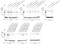
Translational readthrough of amber, opal, and ochre codons by tRNACys-derived suppressor tRNAs. Full-length AQP4-P mRNA containing an amber (A), opal (B) or ochre (C) codon at position 44 (indicated beneath gel) was translated in the presence of the corresponding suppressor tRNA (indicated above gel). Translation was carried out in WG extract in the presence of a Tran35S Label with or without an aptamer. Reactions were analyzed by SDS-PAGE and phosphorimaging. (D) mRNAs described in A were translated in WG extract in the presence of an aptamer and tRNA shown at the top of each lane. Translation reactions were analyzed by SDS-PAGE and phorphorimaging. The identity of the stop codon at residue 44 is indicated below the gel. “–,” Migration of polypeptides that terminate at residue 44; “*” and “↓,” migration of full-length AQP4-P protein.
Importantly, each of the three suppressor tRNAs exhibited a high degree of specificity, since translational readthrough was only observed when the cognate stop codon was present in the mRNA transcript (Fig. 5D). When nonaminoacylated tRNAs were added to translation reactions at similar concentrations, stop codon readthrough was essentially undetectable (data not shown), indicating that these suppressor tRNAs are not appreciably charged with endogenous amino acids in the translation reaction. These results demonstrate that tRNACys-derived suppressor tRNAs can be used to selectively incorporate nonnatural amino acids at all three nonsense codons.
Readthrough of two different nonsense codons in one mRNA using suppressor tRNAs
We next tested whether two different modified amino acids could be concurrently incorporated at two different nonsense codons in a single polypeptide. Yeast [14C]Cys-tRNACysopl was labeled with monobromobimane (MBB) [3-(bromomethyl)-2,5,6-trimethyl-1H,7H-pyrazolo(1,2-α)pyrazole-1,7-dione] and E. coli [3H]Lys-tRNALysamb was labeled with NBD. MBB-Cys-tRNACysopl demonstrated only a slightly lower stability (Table 2) and a slightly lower incorporation efficiency than NBD-Cys-tRNACysopl (Figs. 4B; 5B). Full-length mRNAs containing a UAG at codon 44 and/or a UGA at codon 68 were translated in the presence of ɛNBD-Lys-tRNALysamb, MBB-Cys-tRNACysopl, or both tRNAs as specified in Figure 6. When compared with the translation efficiency of the wild-type (WT) protein that lacked a stop codon, quantification of full-length proteins by phosphorimaging revealed a cumulative readthrough efficiency of 16% at both stop codons (Fig. 6A, cf. lanes 1 and 12). Smaller proteins, ∼5 kDa and 8 kDa in size, were generated by ribosome termination at the stop codons UAG44 and UGA68, respectively. Incorporation of the ɛNBD-lys probe was also confirmed by translating mRNAs truncated at codon 183 [AQP4-P(183)] (Fig. 1B), pelleting the stalled ribosome–nascent chain complexes, and SDS-PAGE. When gels were scanned for NBD1 fluorescence, a clear signal was detected whenever an NBD probe was incorporated (Fig. 6B). Unfortunately, the fluorescent signal of MBB was too low to detect by in-gel scanning (data not shown).
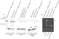
Concurrent incorporation of two modified amino acids at the amber and opal stop codons. (A) Full-length AQP4-P containing L44UAG, L44UGA, or L44UAG plus H68UGA codons (indicated beneath the gel) was translated in WG extract in the presence of a Tran35S Label and an aptamer. ɛNBD-[3H]Lys-tRNALysamb, yeast MBB-[14C]Cys-tRNACysopl, and yeast NBD-[14C]Cys-tRNACysopl were added to the translation reactions as indicated above each lane. Products were analyzed by SDS-PAGE and phosphorimaging. Polypeptides that terminate at residue 44, or readthrough residue 44 but terminate at residue 68, are marked by “–” and “*,” respectively. Full-length polypeptides are marked by “**.” Concurrent readthrough efficiency of both stop codons was 16%, compared with WT protein (cf. lanes 1 and 12). Panels shown are taken from the same gel and exposure time. (B) AQP4-P(183) mRNA containing indicated stop codons (shown at bottom) was translated in WG extract in the presence of aa-tRNAs (shown at top). Ribosome–nascent chain complexes were pelleted, RNase treated, and analyzed by SDS-PAGE. The gel was scanned for NBD fluorescence as described in Materials and Methods.
During these experiments we noticed an interesting relationship between the amounts of protein generated from otherwise equivalent transcripts when stop codons were present in the mRNA. For example, after correcting for methionine content, the number of polypeptides that read through the UAG at codon 44, but terminate at the UGA68 codon (Fig. 6A, lane 10), was actually greater than the number of full-length polypeptides generated in the absence of a stop codon (Fig. 6, lane 1). Note that these proteins contain two and nine Met residues, respectively. Similar results were observed for shorter AQP4-P polypeptides truncated at residue 98 (AQP4-P-98), which also contained the stop codons L44UAG, L44UGA, or L44UAG plus H68UGA (Fig. 7). Again, addition of the cognate tRNAs suppressed termination and resulted in the appearance of appropriate size polypeptide(s) (Fig. 7A).
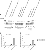
Concurrent readthrough of cognate amber and opal stop codons in a truncated mRNA. (A) AQP4-P(98) mRNAs containing stop codons (shown below the gel) were translated in WG extract containing aa-tRNAs (shown above the gel), and proteins were analyzed by SDS-PAGE following RNase treatment. Polypeptides that terminate at residue 44, or readthrough residue 44 but terminate at residue 68, are marked by “–” and “*,” respectively. Polypeptides that end at residue 98 are shown by “**.” Concurrent readthrough efficiency at both codons was 28% based on phosphorimaging (cf. lanes 1 and 12, **). Polypeptides 44, 68, and 98 residues in length contain two, two, and four methionine residues, respectively. The panels shown are taken from the same gel and exposure time. (B,C) NBD-[3H]Lys-tRNALysamb and yeast MBB-[14C]Cys-tRNACysopl were added individually (B) or together (C) to translation reactions containing apt-12 and programmed with AQP4-P(98) mRNA containing the indicated stop codons (shown below the graphs). The graph shows the amount of [3H]Lys (white bars) or [14C]Cys (gray bars) incorporated into the translated proteins as measured by hot acid precipitable counts. Values show an average of two experiments.
We then quantified the readthrough efficiency of NBD- and MBB-labeled aa-tRNAs by measuring the incorporation of [14C]Cys and [3H]Lys into TCA precipitable protein. Figure 7B shows the amount of probe incorporation when the amber or opal suppressor tRNAs were added individually to separate translation reactions programmed with mRNAs containing UAG, UGA, or both stop codons. Surprisingly, probe incorporation at the upstream UAG codon (ɛNBD-[3H]Lys) increased threefold when translation was terminated at the downstream UGA codon. This was also consistent with the corresponding yields of [35S]Met-labeled polypeptides (Fig. 7A, cf. lanes 3 and 10), which contain four and two Met residues, respectively. Similarly, when both suppressor tRNAs were added together to the translation reactions (Fig. 7C), probe incorporation at the UAG codon was increased by twofold when the downstream UGA codon was present. This most likely occurred because translation termination at the opal codon releases engaged ribosomes and allows them to repeatedly reinitiate translation at the 5′ end of the mRNA. In the absence of the opal codon, ribosomes that readthrough the UAG stop codon stall at the end of the truncated mRNA and thereby reduce the amount of reinitiation. The presence of a downstream (UGA) stop codon therefore allows ribosomes multiple attempts to incorporate the nonnatural amino acid at the upstream UAG codon. Using complementary suppressor tRNAs carrying nonnatural amino acids, together with inhibition of release factors eRF1/eRF3, we achieved a combined readthrough efficiency at two consecutive stop codons of 28% when compared with the amount of parent protein synthesized under identical conditions (Fig. 7A, cf. lanes 1“**” and 12 “**”).
DISCUSSION
In this study, we have developed a generally applicable in vitro system for the site-specific incorporation of multiple probes into one protein. This approach is facile to implement, highly selective for the incorporation site, allows incorporation of diverse probes, and requires no other changes in protein sequence. Cys-tRNAs are excellent candidates to achieve these goals because the reactive sulfhydryl side chain can be conveniently modified with commercially available reagents under mild conditions that leave the labile aminoacyl bond intact. Consistent with this, modified Cys-tRNACys has previously been used to cotranslationally incorporate a fluorescent NBD probe using a eukaryotic cell-free expression system (Alder et al. 2008). However, selective incorporation at the desired site required removal of endogenous native cysteine residues.
To overcome this limitation, we developed tRNACys-derived suppressor tRNAs that selectively suppress translation termination at their cognate stop codons. Radiolabeled and modified suppressor Cys-tRNAs were generated by enzymatic acylation of synthetic tRNA using a combination of E. coli extract and recombinant Cys-aaRS, followed by modification of the sulfhydryl moiety (Komatsoulis and Abelson 1993; Liu et al. 2007a). Although the native cysteine (GCA/ACA) anticodon is thought to be important for recognition by Cys-aaRS (Komatsoulis and Abelson 1993; Liu et al. 2007a), its presence is clearly not essential under the in vitro conditions developed here. However, the E. coli aaRS does not effectively recognize yeast-derived suppressor tRNAs, most probably because two of the enzyme's recognition elements, the G15•G48 Levitt base pair and the GCA anticodon, are missing (Lipman and Hou 1998).
An important, although often overlooked, requirement for cotranslational probe incorporation is that the modified aa-tRNA species must remain stable during the time course of translation. Indeed, we found that [14C]Cys-tRNACys and its suppressor derivatives exhibited low levels of translational incorporation due to rapid deacylation as determined by release of [14C]Cys from acid precipitable tRNA. However, modification of the sulfhydryl moiety with either of the two different fluorescent dyes, MBB and NBD, markedly stabilized the aminoacyl–tRNA bond and increased incorporation efficiency. Based on our observations, the instability of Cys-tRNAs seems to arise from both a chemical component, most likely caused by the reactive sulfhydryl side chain as demonstrated by spontaneous deacylation in buffer (Strickland and Jacobson 1972), and an enzymatic component, most probably due to trans-editing aaRS activity present in both RRL and the WG extract (Jakubowski and Goldman 1992; Ahel et al. 2003; Ibba and Söll 2004). Cysteine modification markedly improved the chemical stability of Cys-tRNA and overcame trans-editing activity in the WG extract, but had little stabilizing effect in RRL. The contribution of aa-tRNA stability to stop codon suppression is therefore dependent upon both the nature of the attached aminoacyl moiety and the relevant deacylation enzymes present in the translation system (Ahel et al. 2003).
Interestingly, NBD-Cys-tRNACysamb enabled translational readthrough in the WG extract with an efficiency that approached ɛNBD-Lys-tRNALysamb. Thus, both amber suppressor tRNAs compete effectively with the eukaryotic release factor eRF1/eRF3, even though they are derived from different tRNA scaffolds. The readthrough at the opal and ochre codons, however, was markedly less efficient. A similar pattern obtained for orthogonal tRNAGln-based suppressors was previously attributed to differences in aminoacylation efficiency (Köhrer et al. 2004). Because the aa-tRNACys-derived suppressors used here are all present at equivalent concentrations, our results suggest that reduced suppression at the opal and ochre codons may also be caused by less efficient competition with translation termination factor eRF1/eRF3 (Köhrer et al. 2004; Taira et al. 2005; Rodriguez et al. 2007). One possible explanation is that Watson–Crick codon–anticodon base-pairing with amber, as opposed to opal and ochre, tRNAs in the ribosome A site more favorably stimulate the forward reaction of eEF-1α GTP hydrolysis and peptide bond formation (Rodnina et al. 2005; Youngman et al. 2008). Weaker ternary interactions between mRNA, tRNA, and the ribosome might therefore lead to a premature aa-tRNA release, possibly necessitating multiple tRNA entry events, each of which would presumably compete with the release factors for translational readthrough. Although in vitro-synthesized tRNACysamb, tRNACysopl, and tRNACysoch lack normal base modifications, they are identical in every aspect with the exception of the anticodon. Thus, unless the anticodon bases or subtle differences in their hydrogen bonding to the amber, opal, and ochre codons exert long-range structural effects, these tRNAs would not be expected to exhibit major conformational differences at the ribosome decoding center.
If the rate of tRNA entry into the ribosome A site and subsequent induced fit are similar for these matched tRNACys-derived suppressors, then differences in suppression efficiency observed here likely reflect intrinsic differences in the binding of eRF1/eRF3 to amber, opal, and ochre stop codons, and/or subsequent conformational changes required to form the mature termination complex (Kisselev and Buckingham 2000; Salas-Marco and Bedwell 2004; Youngman et al. 2008). The observation that an aptamer preferentially stimulates readthrough at opal and ochre codons to a greater extent than at amber codons, supports this notion and suggests that eRF1/eRF3 retains a hierarchy in stop codon recognition, i.e., ochre > opal > amber. This behavior resembles the selective stop codon reprogramming observed in ciliates (Chavatte et al. 2003; Salas-Marco et al. 2006), even though all three codons are utilized for translation termination in higher eukaryotes. Given that little is known about the relative binding affinities and molecular details of how eRF1/eRF3 induces translation termination (Youngman et al. 2008), the set of tRNAs developed here may prove useful in deciphering mechanisms that underlie differences in suppression and/or termination efficiency.
The concurrent incorporation of two nonnative amino acids obtained here using modified aminoacyl amber and opal tRNAs was approximately 10 times higher for truncated mRNA and six times higher for full-length mRNA than has been previously observed for native amino acids in a eukaryotic expression system (Köhrer et al. 2004). Important and novel aspects of our approach include: (1) improved conditions for in vitro aminoacylation of modified tRNACys; (2) selective incorporation of nonnative amino acids at all three cognate stop codons; (3) improved readthrough efficiency obtained by aptamer-induced inhibition of eukaryotic release factors; and (4) the ability to accurately quantify translation readthrough at both probe sites by 14C and 3H isotope incorporation.
Somewhat surprisingly, the presence of a second engineered stop codon substantially improved the apparent readthrough efficiency at the first stop codon. The readthrough efficiency is conventionally defined as the fraction of translating ribosomes that bypass the stop codon and continue translating downstream coding sequence. In most in vivo systems, however, it is difficult to quantify polypeptides that terminate prematurely, and as a result, the apparent readthrough efficiency is frequently determined by comparing the amount of protein generated from mRNAs that lack (or contain) a stop codon. This is a reasonable assessment because the yield of protein containing the nonnative amino acid is the most important parameter for most biological applications. Interestingly, our analysis indicates that translation termination at a second, C-terminal (UGA) stop codon enables ribosomes to rapidly reinitiate translation, thereby increasing the number of chances for readthrough at the first (UAG) stop codon. In this manner ribosome “recycling” can actually increase the “apparent” UAG codon readthrough efficiency to greater than 100%, as shown in Figure 7. By extension, recycling also increases the number of ribosomes that reach and read through the second (UGA) codon. Because the magnitude of this increase is dependent upon the time it takes for the ribosome to finish translation, concurrent readthrough will, to some extent, be affected by the distance between upstream and downstream stop codons, the site of truncation, and/or the length of the translated polypeptide. Optimization of coding sequence length and stop codon positions are therefore important considerations. Readthrough efficiency was also increased for mRNA truncated after the final (endogenous) stop codon when compared with mRNA transcribed from supercoiled plasmid (data not shown), suggesting that the effects of the 3′ untranslated region on translation initiation play an important role. Because these observations are made using an in vitro expression system, additional work is needed to determine whether similar mechanisms affect sequential stop codon readthrough in intact cells (Köhrer et al. 2004).
In conclusion, the development of tRNACys-derived suppressor tRNAs now allows simultaneous incorporation of two nonnatural amino acids in the same protein at efficiencies suitable for diverse biochemical and biophysical studies. The strategy described here provides a promising new technology to position multiple probes at defined locations in a protein with other minimal alterations of its natural sequence.
MATERIALS AND METHODS
Plasmids and transcription
Plasmid pSP64-MIWC2 (Shi et al. 1995), here called AQP4-P to conform with new nomenclature, encodes the 46 N-terminal residues of AQP4, a Thr-Val linker (BsteII restriction site), and the 142 C-terminal residues (amino acids 88–229) of bovine preprolactin (pPL) (diagrammed in Fig. 1A). Codon Leu44 of AQP4-P was converted to TGC (Cys), TAG (amber), TGA (opal), or TAA (ochre) by PCR overlap extension (Ho et al. 1989). Similarly, Cys49 was converted to alanine, and, where indicated, His68 was converted to TGA (opal). The sequence context of engineered stop codons, which is an important parameter for readthrough efficiency, is AACCCCXXXCCCGTG and ACCCACXXXGAAGTA for residue 44 and residue 68, respectively, where “XXX” represents the introduced stop codon. cDNA was truncated by PCR amplification using a 5′-oligonucleotide complementary to the pSP64 vector (base pair 2757) and a 3′-oligonucleotide ending at codon 98 or 183, which converted the last translated codon to valine to increase peptidyl-tRNA bond stability. Plasmid DNA or a truncated PCR product was transcribed in vitro using SP6 polymerase under standard conditions described elsewhere (Oberdorf and Skach 2002).
Plasmids encoding yeast tRNACys or E. coli tRNALysamb are described elsewhere (Flanagan et al. 2003; Alder et al. 2008). Plasmids encoding E. coli and human tRNACys and their corresponding aaRS (Liu et al. 2007a) were kindly provided by Ya-Ming Hou (Thomas Jefferson University). Except for human tRNACys, all tRNA sequences together with the T7 promoter were ligated into the SP64 plasmid by using EcoRI and HindIII sites introduced by PCR. The cysteine anticodon was then converted to an amber, opal, or ochre anticodon by PCR overlap extension. For transcription, tRNA sequences and the T7 promoter were amplified by PCR, and the resulting DNA transcribed in vitro using T7 RNA polymerase. The tRNA transcripts were purified by FPLC using a MonoQ column as described previously (Flanagan et al. 2003).
The RNA aptamer was generated from synthetic overlapping DNA oligonucleotides encoding aptamer-12 (Carnes et al. 2000), T7 promoter, and EcoRI and HindIII sites. The PCR products were ligated into pSP64, and transcription and aptamer purification were performed as for tRNAs.
tRNA aminoacylation and modification of the amino acid side chain
tRNA aminoacylation and purification were performed as described (Johnson and Slobin 1980; Alder et al. 2008) with the following modifications: For tRNALysamb, reactions contained 100 mM HEPES (pH 8.0), 8 mM MgCl2, 1 mM DTT, 4 mM ATP, 0.1 mM CTP, 50 A260 units of purifed tRNALysamb, 25% (v/v) DMSO, 12 μM [14C]Lys (Sigma) or [3H]Lys (GE Healthcare), and 10% (v/v) S-100 E. coli enzyme extract (Menninger et al. 1970; Johnson et al. 1976; Alder et al. 2008). The reaction was incubated at 37°C for 90 min. For tRNACys and the suppressor derivatives, the reactions contained 100 mM HEPES (pH 7.5), 10 mM MgCl2, 10 mM DTT, 4 mM ATP, 0.1 mM CTP, 50 A260 units of tRNA, 25% (v/v) DMSO, 5 μM [14C]cystine (Perkin Elmer), preincubated with DTT, and 10% (v/v) of S-100 E. coli enzyme extract as previously described (Crowley et al. 1993). Where indicated, His6-tagged E. coli or human Cys-aaRS was expressed in E. coli, purified as previously described (Liu et al. 2007a), and added to the final concentrations of 125 and 320 μg/mL, respectively, together with 6% (v/v) S-100 extract. The reaction was carried out at 37°C for 45 min.
The ɛ-amino group of Lys-tRNALysamb was chemically modified with succinimidyl 6-[N-(7-nitrobenz-2-oxa-1,3-diazol-4-yl)amino]hexanoate (Invitrogen) (Crowley et al. 1993). The thiol group of Cys-tRNAs was chemically modified with N,N′-dimethyl-N-(iodoacetyl)-N′-(7-nitrobenz-2-oxa-1,3-diazol-4-yl)ethylenediamine (Invitrogen) as previuosly described (Alder et al. 2008). Alternatively, 3-(bromomethyl)-2,5,6-trimethyl-1H,7H-Pyrazolo(1,2-α)pyrazole-1,7-dione (Invitrogen) was coupled to Cys-tRNAs under the same conditions. All modified aa-tRNAs were purified by reversed phase-HPLC (Alder et al. 2008).
In vitro translation
AQP4-P fusion proteins were translated at 24°C for 1 h in 10 μL cell-free translation reactions containing 20% (v/v) of transcription reaction and 40% (v/v) hemin-supplemented RRL (Oberdorf and Skach 2002) or 20% (v/v) WG extract (Erickson and Blobel 1983). For undesalted RRL, 10 mM Tris acetate (pH 7.5), 100 mM KOAc, and 2 mM MgOAc were added, while desalted WG extract was adjusted to a final concentration of 20 mM HEPES/KOH (pH 7.5), 140 mM KOAc, and 3 mM MgOAc. Translation mixtures also contained: 2 mM DTT, 0.4 mM spermidine, 1 mM ATP, 1 mM GTP, 12 mM creatine phosphate, 40 μM of all 20 amino acids, 40 μg/mL creatine kinase, 0.2 U/μL RNase inhibitor, and 1 μM of tRNA, as indicated. Where Cys-tRNACys was added, cysteine was omitted from the translation mixture. To detect translation products by SDS-PAGE, 1 μCi/μL Tran35S Label (MP Biomedicals) was added instead of methionine. Where indicated, 1 μM of the RNA aptamer was added. For stability measurements of aminoacyl-tRNAs, a mock transcription mixture lacking a DNA template, was used.
Analysis of translation products
To quantify incorporation of 14C-labeled or 3H-labeled amino acids, 4 μL of translation reaction was incubated in 96 μL 1 M NaOH, 2% (v/v) H2O2 at 37°C for 10 min, and protein was precipitated in 10% (w/v) TCA at 85°C (Daniel et al. 2008). Precipitate was collected by filtration (Durapore HVLP02500, Millipore) and subjected to liquid scintillation counting (Beckmann 6500). Background isotope incorporation was determined using an mRNA construct lacking the codon for incorporation. To measure aminoacyl-tRNA stability, aliquots were taken after 0-, 1-, 5-, 15-, and 60-min incubations and analyzed as above, except that bleaching was not performed, and precipitation was carried out on ice for 30 min to prevent aminoacyl-tRNA hydrolysis. To distinguish between isotopes, samples that contained only 3H or 14C were used to set energy windows in the scintillation counter. After background subtraction, the lower energy window detected 100% of the 3H counts (counting efficiency of 9.6% cpm/dpm) and 33% of the 14C counts (counting efficiency of 85% cpm/dpm), whereas 67% of the 14C counts were detected in the higher energy window. Since all samples were treated similarly, quenching of the samples, and therefore the energy distribution of the counts, remained constant (data not shown). By subtracting the relative contribution of 14C counts from the lower energy window and adding to the higher energy window net 3H and 14C counts were determined for samples containing both radioisotopes.
For SDS-PAGE, 1 μL of 35S-containing translation was separated on 12%–17% (w/v) polacrylamide gels. Gels were dried, exposed on a phosphorimaging screen (Eastman Kodak), and analyzed using a Bio-Rad FX molecular imager with Quantity One software (Bio-Rad). For in-gel fluorescence detection, ribosome–nascent chain complexes were pelleted at 350,000 g for 1 h at 4°C and resuspended in 10 mM Tris-HCl (pH 8.0) and 0.1% (w/v) SDS. Peptidyl tRNA was digested with 0.05 mg/mL RNase A at 24°C for 15 min, and the samples were analyzed by SDS-PAGE. In-gel fluorescence was detected using a Fuji film FLA-5000 imager equipped with a 473 nm laser and a >510-nm high-pass filter.
ACKNOWLEDGMENTS
This work was supported by a Netherlands Organization for Scientific Research (NWO) grant (J.G.), Cystic Fibrosis Foundation Therapeutics grant SKACH05X0 (W.R.S.), and National Institutes of Health grants DK51818 and GM53457 (W.R.S.). We thank Ya-Ming Hou for generously providing plasmids encoding E. coli tRNACys, human tRNACys, and E. coli, and human-derived aaRS. J.G., S.J.K., and W.R.S. designed the experiments, and analyzed the data. J.G., S.J.K., and Z.Y. performed the experiments. J.G. and W.R.S. wrote the manuscript. A.E.J. provided materials and advice on the manuscript.
Footnotes
-
Reprint request to: William R. Skach, Department of Biochemistry and Molecular Biology, Oregon Health and Sciences University, 3181 SW Sam Jackson Park Road, L-224, Portland, OR 97231, USA; e-mail: skachw{at}ohsu.edu; fax: (503) 494-8393.
-
Article published online ahead of print. Article and publication date are at http://www.rnajournal.org/cgi/doi/10.1261/rna.2024810.
- Received December 3, 2009.
- Accepted May 12, 2010.
- Copyright © 2010 RNA Society




