


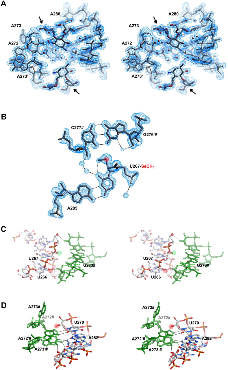
HIV-1 DIS RNA/drug complex (Freisz et al. 2008). (A) Stereo view of the experimental MAD electron density map. The map clearly shows the bulge region (A272, A273, A280) of RNA, two ribostamycin aminoglycosides (indicated with black arrows), and surrounding water molecules (red spheres). (B) View of the U267-SeCH3 A285′ base-pair in the HIV DIS structure at 1.5 Å resolution. Se atom is depicted by a pink sphere. 2Fo-Fc electron density map is represented around the model. The C277-G276′ base-pair from a symmetry-related molecule is also shown. Hydrogen bonds and the intraresidue C-H(methyl)-O bond in the modified uridine are represented with black and gray dotted lines, respectively. (C, D) Stereo views showing crystal packing interactions around U266, U267 (C), and U270 (D) residues. Symmetry-related molecules are shown in green, and nucleotides are indicated with a hash mark (#). 2′-OH groups of U266 and U270, which do not allow 2′-SeCH3 modifications, are shown with a pink circle. The 2′-OH group of U267, which can accommodate 2′-SeCH3 modification, is depicted with a green circle. The hydroxyl of U266 interacts with a 2′-OH group from a symmetry-related molecule (C), and the hydroxyl of U270 is involved in an A minor interaction with a bulged-out A272′# (D), whereas 2′-OH of U267 is not H-bonded and available for 2′-SeCH3 modification (C). Residues U266′, U267′, and U270′ from the second molecule in the asymmetric unit are not involved into crystal packing (not shown).




