


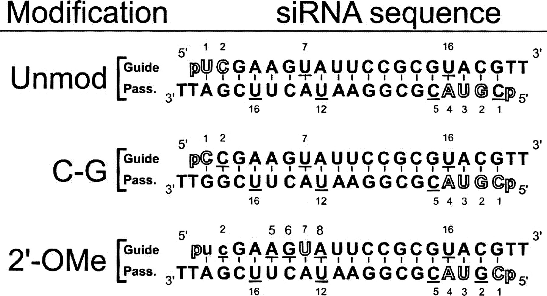
siRNAs used in this study as derived from Martinez et al. (2002), with guide and passenger strands indicated. All three constructs carry 5′-phosphates as well as two deoxythymidines on the 3′-end of each strand. In the C–G siRNA, a C–G base pair replaces the closing U–A base pair at the 5′-guide strand terminus. The 2′-O-Me siRNA harbors two 2′-O-Me modifications at positions 1 and 2 of the guide strand (depicted in lowercase letters). An outlined nucleotide indicates a nucleolytic hit 3′ to that position and represents ≥5% of the total lane intensity, while an underlined nucleotide indicates a hit 3′ to that position that is >10-fold of background, but <5% of the total lane intensity.




