


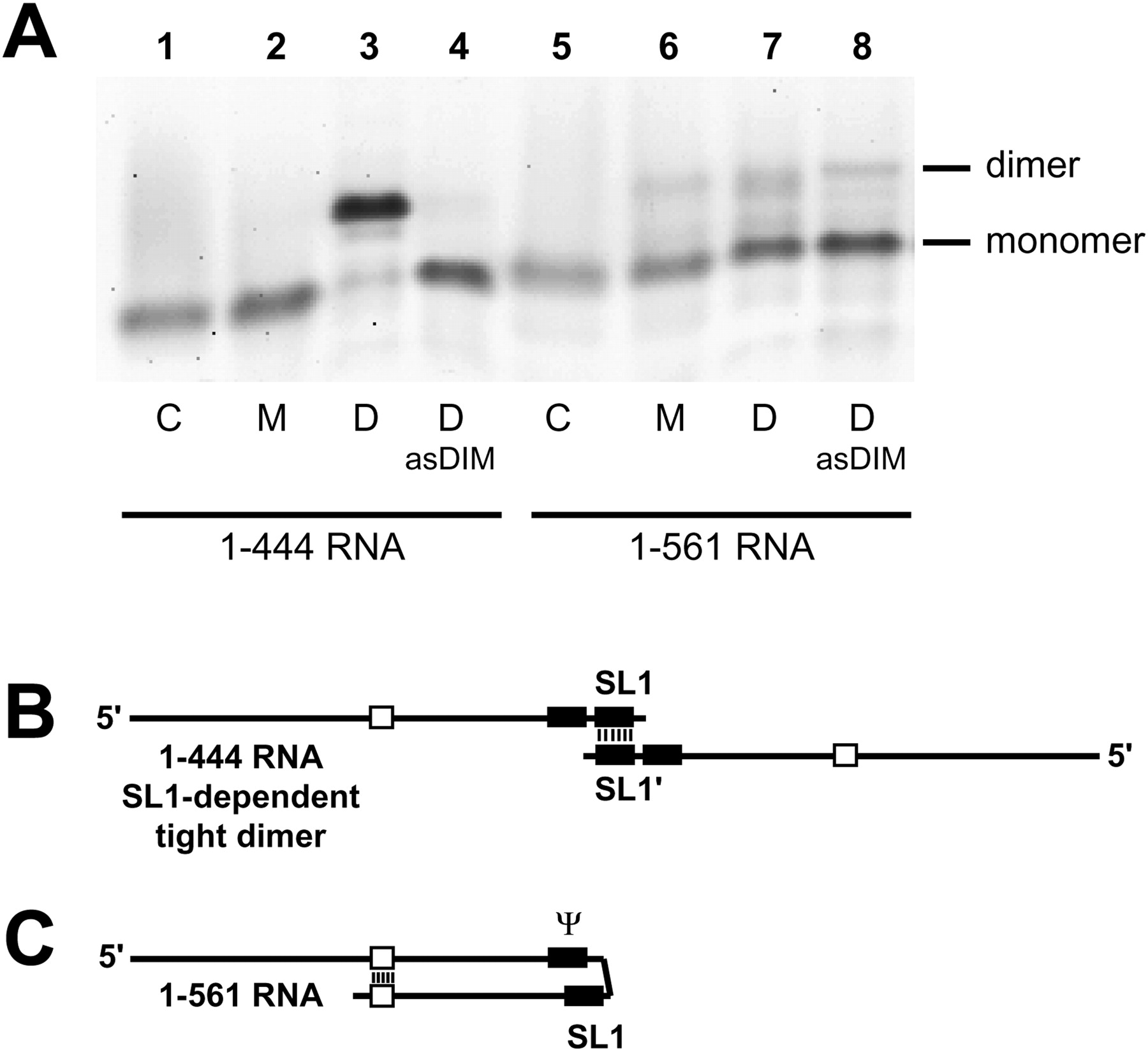
TBE-resistant dimerization of 1–444 and 1–561 RNAs. (A) 1–444 and 1–561 RNAs were assayed for dimerization at 55°C with monomer (M) or dimer (D) buffer. Dimerization was also assayed in the presence of a 20-fold excess of antisense DNA oligonucleotide asDIM, which is complementary to nucleotides 397–426. After incubation for 30 min, samples were subjected to electrophoresis on a TBE agarose gel at 28°C. Only tight dimers withstand the warm TBE electrophoresis. (Control lanes C) Monomeric RNA that was denatured at 90°C, then quenched on ice immediately prior to loading. (B) Schematic representation of the tight dimer of 1–444 RNA from A, lane 3. The two molecules interact through an extended base pairing of the SL1 elements. (C) Schematic representation of the proposed monomeric form of 1–561 RNA from A, lane 7. Upon incubation of 1–561 RNA at high temperature, there is an intramolecular folding of SL1 that competes with the intermolecular interaction (i.e., dimerization). The SL1 structure is shown partially base paired to the upstream encapsidation signal Ψ, as described in Lanchy et al. (2003).




