


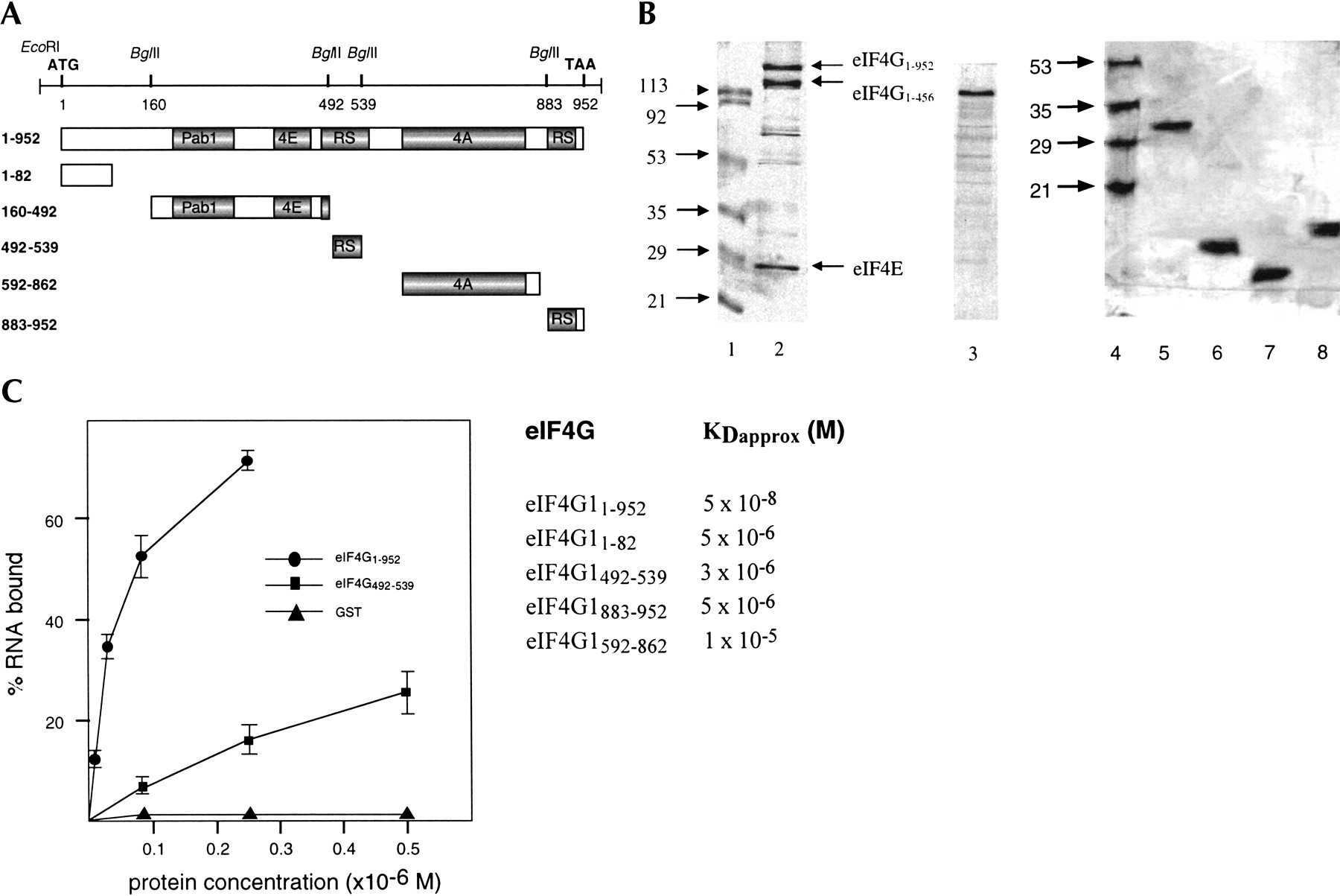
Identification of eIF4G1 RNA-binding sites. (A) Schematic representation of yeast eIF4G1 and eIF4G1 fragments. The DNA with the relevant restriction sites used for subcloning is shown on top. ATG and TAA indicate start and end of the open reading frame. The numbers indicate amino acid positions. Pab1, poly(A)-binding protein 1 binding domain; 4E, eIF4E-binding domain; RS, arginine- and serine-rich domain; 4A, eIF4A-binding domain. (B) About 1–2 μg of each protein were fractionated on 15% (lanes 1–3) and 20% (lanes 4–8) SDS polyacrylamide gels and stained with Coomassie blue. (Lanes 1,4) molecular weight markers (numbers indicate kilodaltons), (lane 2) eIF4G11–952, (lane 3) eIF4G1160–492, (lane 5) eIF4G1592–862, (lane 6) eIF4G1883–952, (lane 7) eIF4G1492–539, (lane 8) eIF4G11–82. (C) Filter-binding assay. On the left, the titration curves of three of the proteins are shown. Proteins were titrated with 0.25 pmole [32P]-labeled 38-nt RNA in a 20-μL reaction volume containing buffer A. On the right, KDapprox. values for eIF4G1 proteins are shown. For estimation of protein concentration of full-length eIF4G1, only the slowest migrating band (B, lane 3) was included.




