


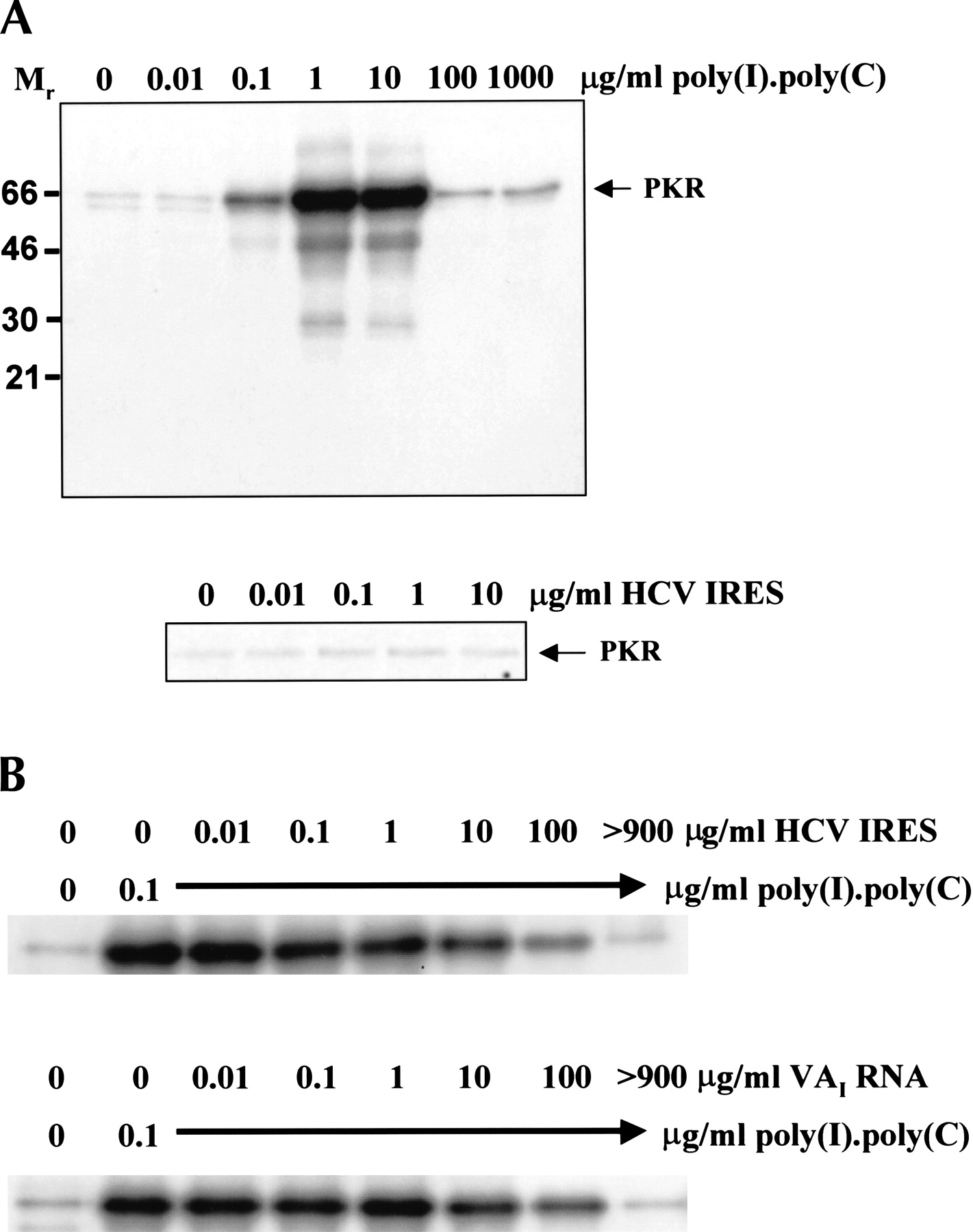
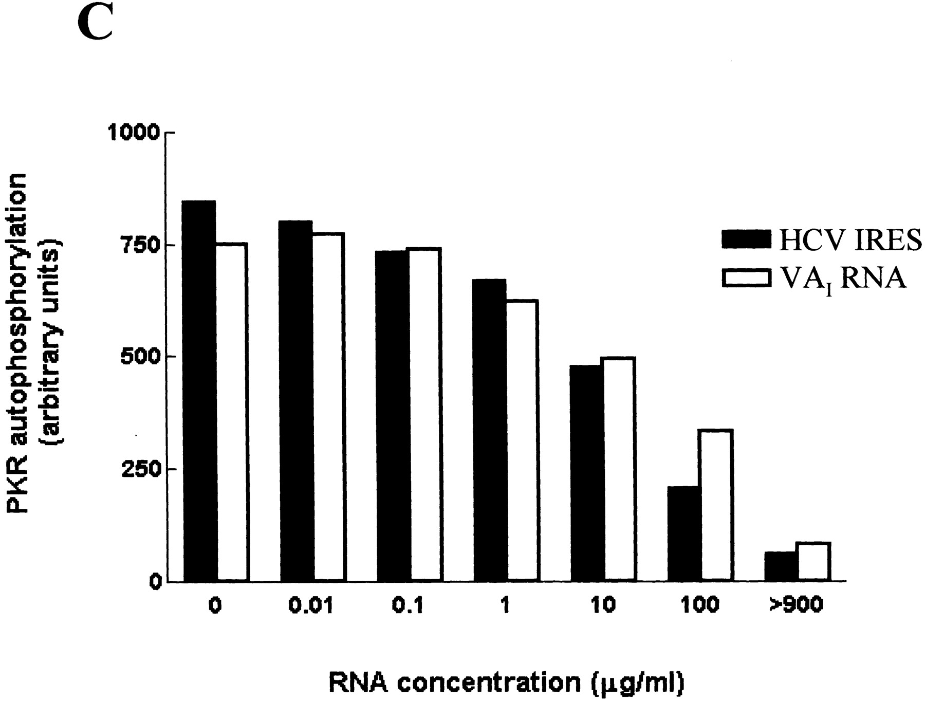
Effects of the HCV IRES on the protein kinase activity of PKR. (A) Purified wild-type PKR was incubated with the indicated amounts of poly(I).poly(C) (upper panel) or the HCV IRES transcript (lower panel) in the presence of [γ32P]ATP, as described in Materials and Methods. The extent of activation of the protein kinase, as reflected in the level of autophosphorylation, was determined by SDS gel electrophoresis and phosphorimaging. The positions of molecular mass markers and PKR are indicated. (B) PKR was incubated with poly(I).poly(C) (0.1 μg/mL) in the presence of the indicated concentrations of the HCV IRES transcript (upper panel) or adenovirus VAI RNA (lower panel) and the extent of autophosphorylation of the protein kinase was determined as in A. (C) The dose-responses for the inhibitory effects of the HCV IRES and VAI RNA on PKR autophosphorylation, as determined by phosphorimaging analysis of the data in B, are depicted graphically.




