


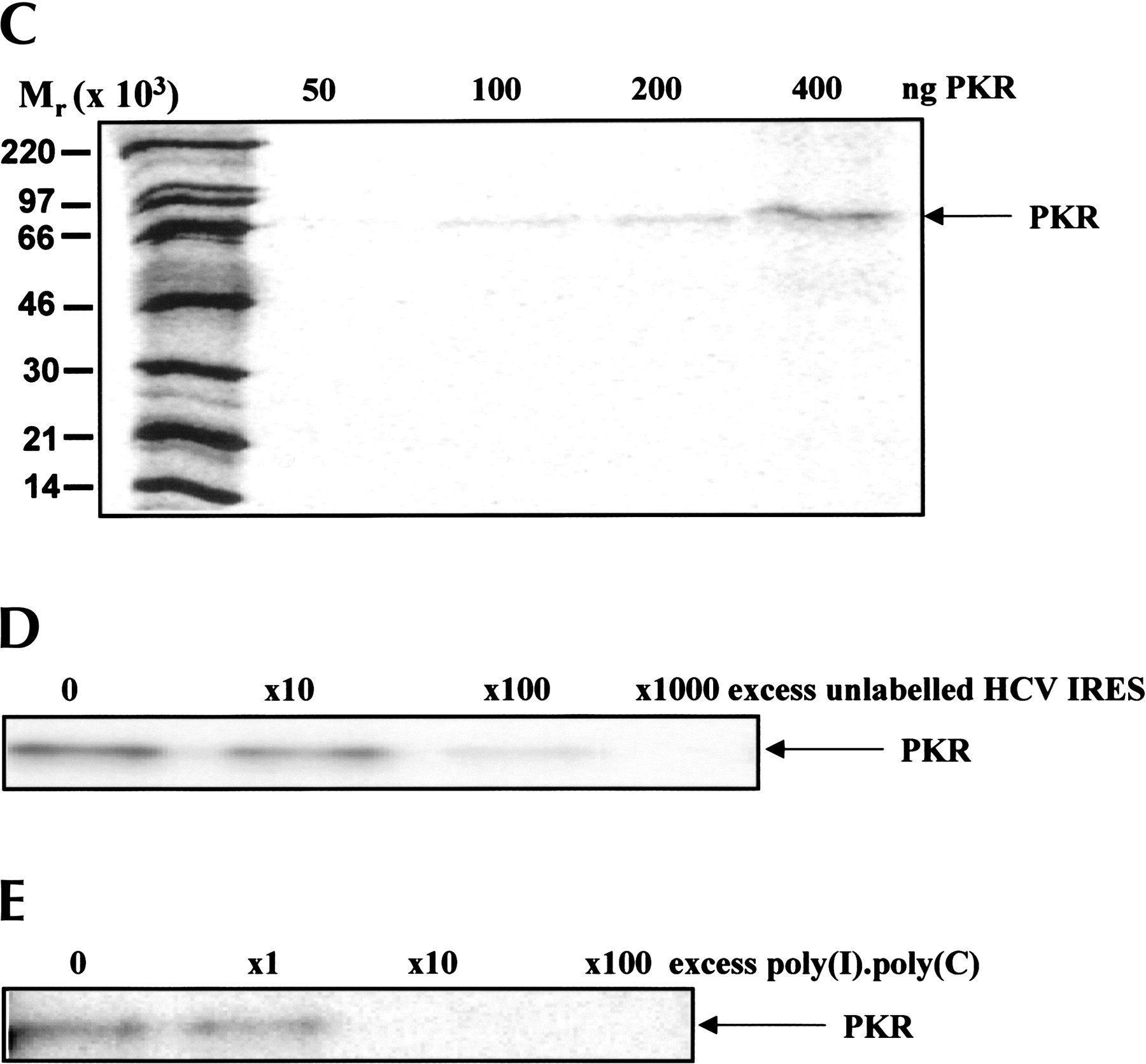
Interaction of the HCV IRES with PKR. The HCV IRES sequence was labeled in vitro by transcription in the presence of [α32P]UTP as described in Materials and Methods. (A) The radioactive transcript (105 cpm) was incubated for 15 min at 30°C with the indicated amounts of purified PKR K296R and the extent of complex formation was analysed by filter binding and phosphorimaging as described in Materials and Methods. (B) Incubations were carried out as in Panel A, except that the indicated amounts of unlabeled poly(I).poly(C) were included as competitor for the labeled IRES. (C) Incubations were carried out as in Panel A and the RNA–protein complexes were then subjected to UV cross-linking followed by digestion with RNases A and T1. The labeled protein–RNA complexes were then analyzed by SDS gel electrophoresis and phosphorimaging. Molecular mass markers and the position of migration of PKR are indicated. (D) A similar experiment to that described in C was performed except that increasing amounts of unlabeled HCV IRES transcript were included in the incubations, as shown. (E) The radiolabeled HCV IRES was incubated with PKR in the presence of the indicated amounts of unlabeled poly(I).poly(C), followed by UV cross-linking and gel analysis as in C and D. The molar excess levels of poly(I).poly(C) over the HCV IRES were calculated on the basis of an average Mr of 2 × 10⁵ for the double-stranded polymer.




