


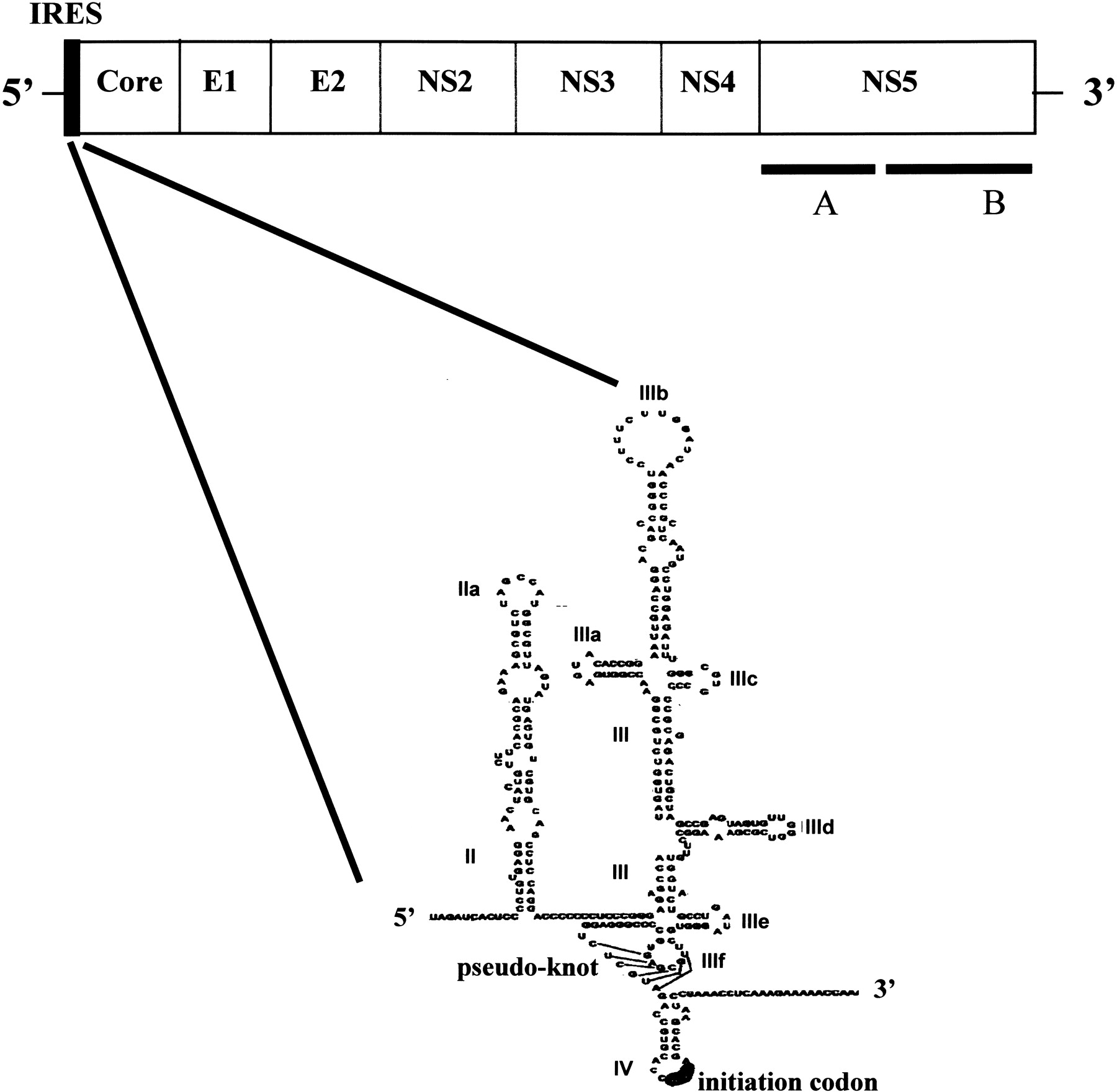
The hepatitis C virus genome and structure of the IRES element. A schematic representation of the organization of the HCV genome is shown at the top with the positions of the IRES and coding regions for the structural proteins (core, E1, and E2) and nonstructural proteins (NS2, NS3, NS4, NS5A, and NS5B) indicated. The IRES structure is shown below. The regions of secondary structure, the numbering of domains II–IV, and the positions of the pseudoknot and initiation codon are as described in Wang et al. (2000), from which this diagram has been modified. The elements of extensively base-paired and/or tertiary structure may provide a basis for recognition by eIF3, 40S ribosomal subunits, and a variety of cellular proteins, including PKR, as described in the text.




