


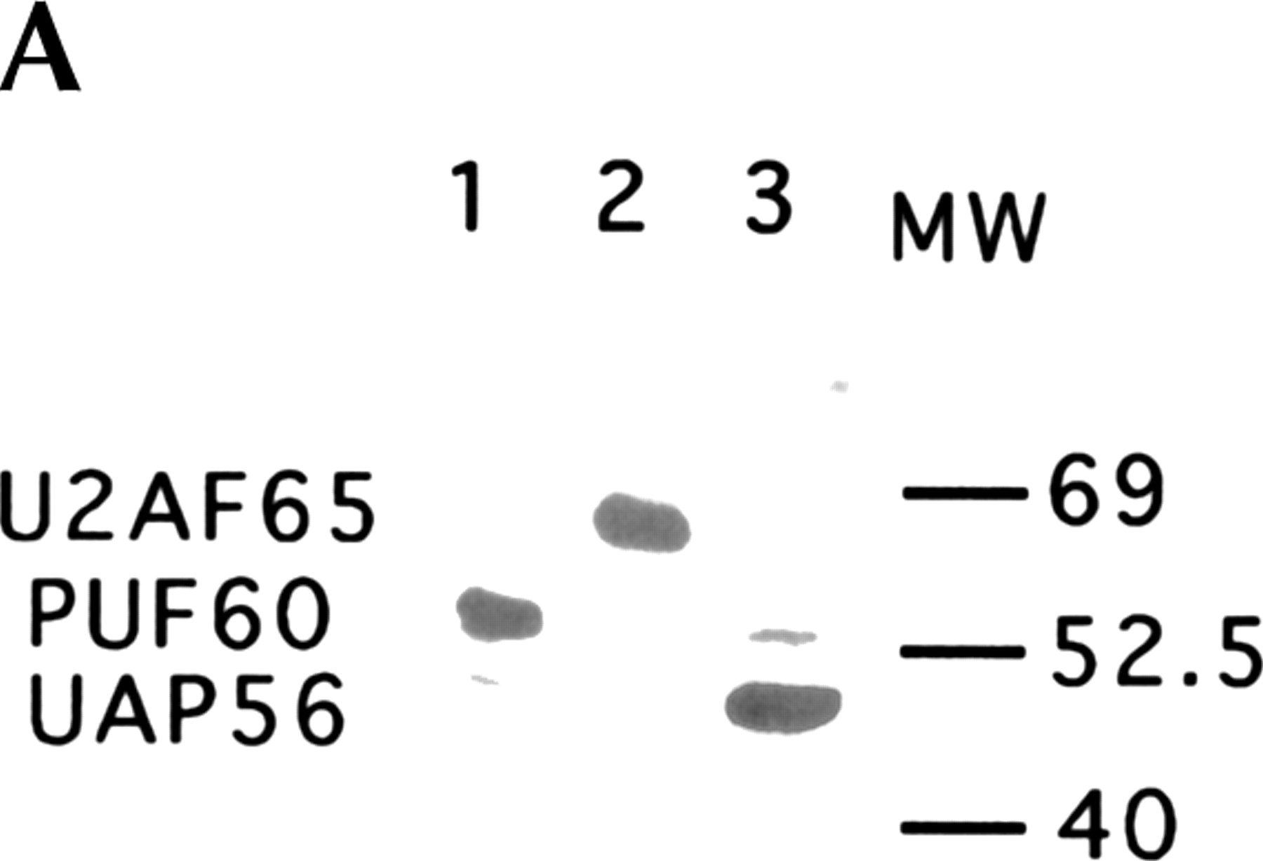
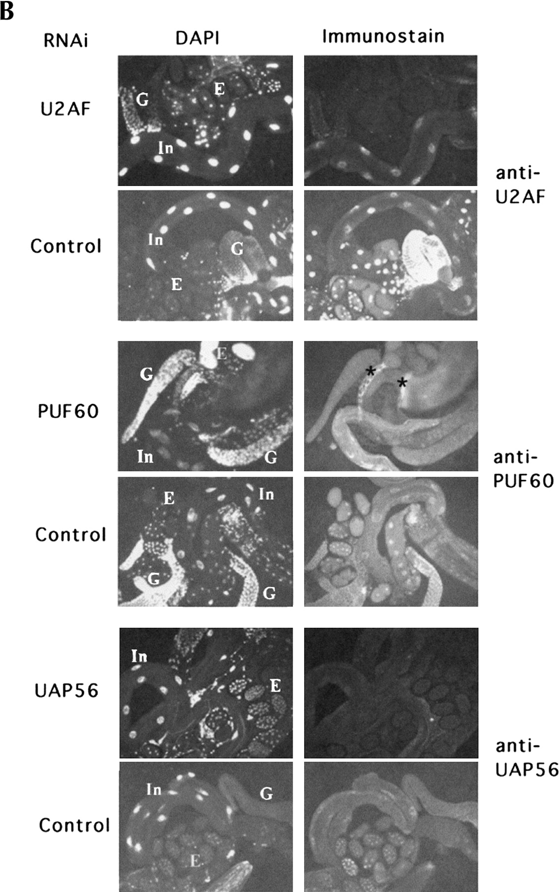
RNAi of UAP56, PUF60, and U2AF65 effectively reduces protein levels. (A) Western blot showing the specificity of the three antibodies. In each panel, the same amount of embryonic extract was electrophoresed, and the blotted protein was reacted with a single antibody: (1) anti-PUF60, (2) anti-U2AF65, or (3) anti-UAP56. (B) Animals were injected with dsRNA of U2AF65, PUF60, or UAP56. The control specimens were uninjected age-paired worms from the pool of animals used for injection. Worms were immunostained with antibodies to the proteins that were subjected to RNAi. Paired DAPI-staining (nuclei) images are shown for each specimen. Large DAPI-staining nuclei characterize intestinal cells (In). Gonads (G) have many close-packed nuclei, and oocytes and embryos (E) have distinct nuclei. Staining for each of the antibodies is predominantly nuclear. One specimen (RNAi of PUF60) has bright artifactual staining (☆) that is extracellular staining of the spermatheca and the edge of the cuticle.




