


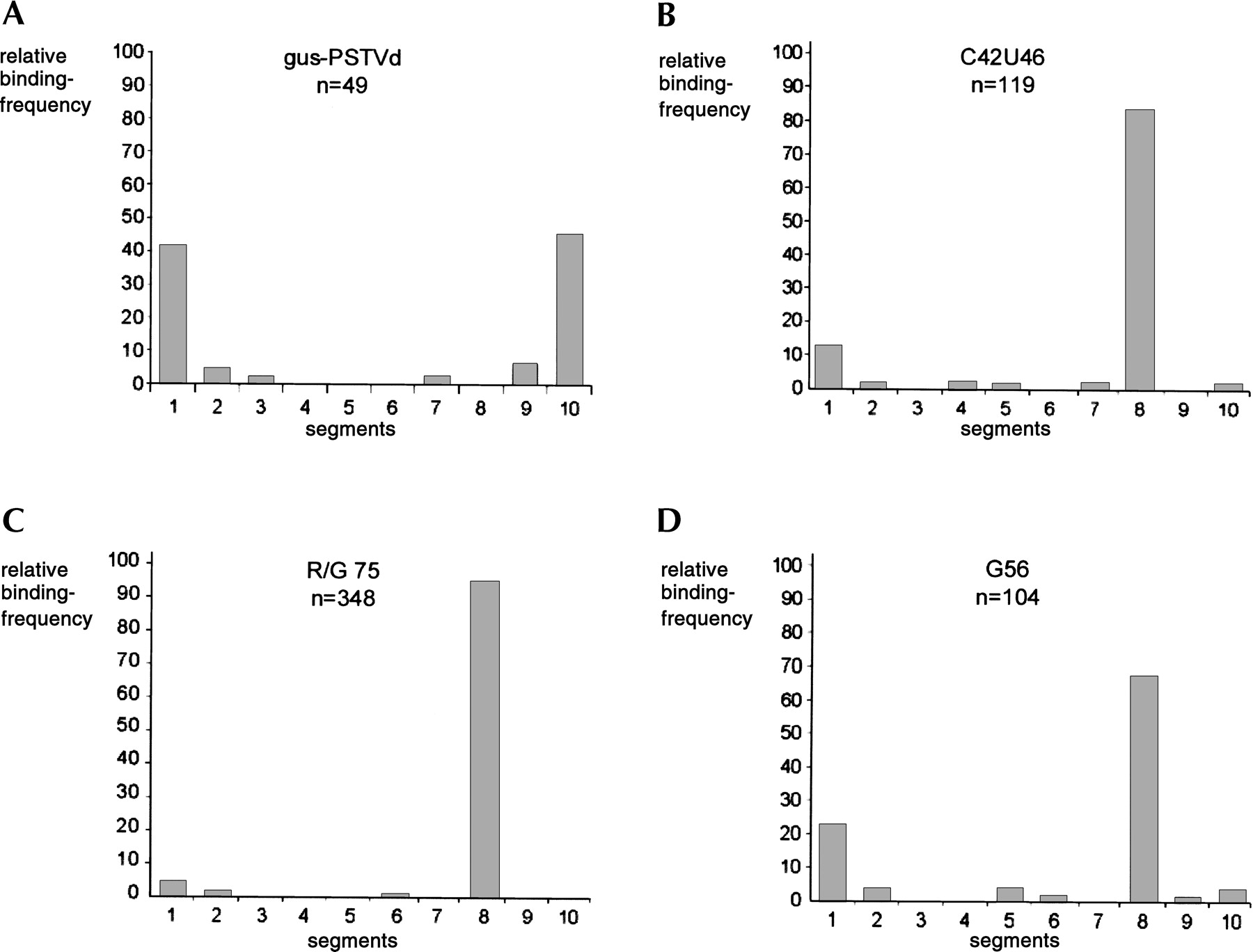
Distribution of ADAR2 binding on the different target molecules. (A) Binding to the gus-PSTVd backbone RNA. The orientation of the molecule was arbitrarily determined (see Materials and Methods), therefore the distribution of binding appears symmetrical. ADAR2 binding to the backbone (B) with the R/G 75 (wild-type) stem-loop inserted, (C) with the C42U46 stem-loop inserted, and (D) with the G56 point mutation stem-loop inserted. (B–D) The orientation was unambiguously determined by the bifurcation on the right end of the molecule (Fig. 1). Bound protein was assigned to one of 10 segments (numbered from left to right) of the RNA molecule. Except for two cases, only one protein was bound to the RNA. (n) Number of RNA molecules examined.




