


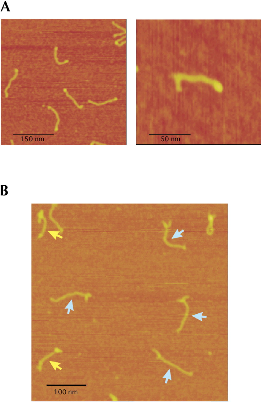
FIGURE 3.
SFM images of ADAR2 binding to different substrates. (A) SFM image of gus-PSTVd backbone molecules with bound ADAR2 proteins. (Right panel) A single molecule is enlarged. Protein binding can be readily seen on the right end of the RNA. (B) SFM image of gus-PSTVd-R/G75 molecules with bound ADAR2 proteins. Note the bifurcation of the RNA, which was used to determine the orientation of the molecule. Yellow arrows indicate RNAs with ADAR2 bound to the editing stem-loop; blue arrows indicate RNAs without bound ADAR2. Note that in all images the background of unbound protein is very low. Images show selected frames.




