


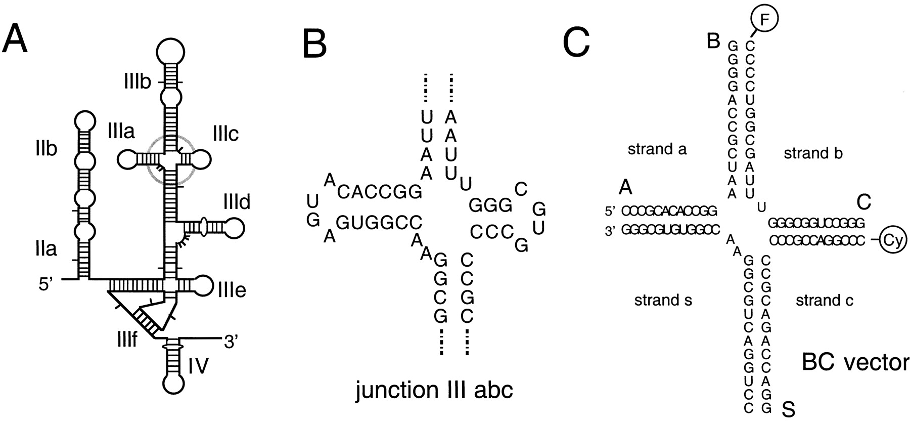
Junction IIIabc of the HCV IRES, and its global conformation. (A) Schematic of the secondary structure of the IRES, with junction IIIabc circled. (B) The local sequence of the junction IIIabc. For the conformational analysis, we have extended the arms, and also created a perfect 4H junction by removal of the unpaired nucleotides. (C) The sequence of the junction used in FRET experiments. All four arms comprise 12 bp with no imperfections, with the natural sequence preserved to the maximum distance in each case – the same central sequences were used in the electrophoresis experiments. We denote the helical arms in both junctions as A, B, C, and S, corresponding to the helices of stem loops III a–c, and the main domain III helix [here called S (= starred) to be consistent with the use of helix III* in Keift et al. (2002)] in the IRES RNA. FRET vectors are named according to the helical arms carrying the fluorescein and Cy3 fluorophores, in that order; the example shown is the BC vector.




