


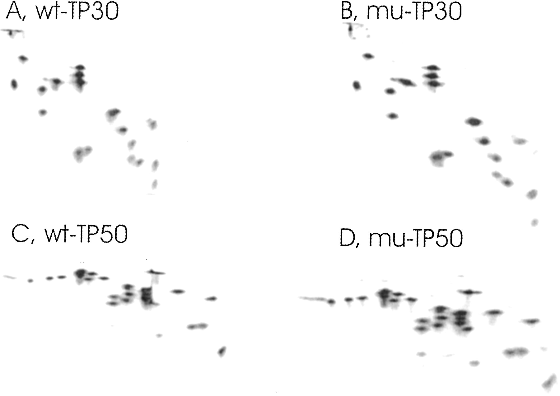
FIGURE 3.
Two-dimensional polyacrylamide gel electrophoresis of 50S and 30S ribosomal proteins from the dnaK+ (MC41.14) and ΔdnaK (BB11) strains. TP50 and TP30 were prepared and subjected to two-dimensional polyacrylamide gel electrophoresis according to Geyl et al. (1981). (A,B) TP30 from 30S subunits of the MC41.14 dnaK+ strain (wt-TP30) and mutant BB11 (ΔdnaK) strain (mu-TP30), respectively. (C,D) TP50 from 50S subunits of the MC41.14 dnaK+ strain (wt-TP50) and mutant BB11 (ΔdnaK) strain (mu-TP50), respectively.




