


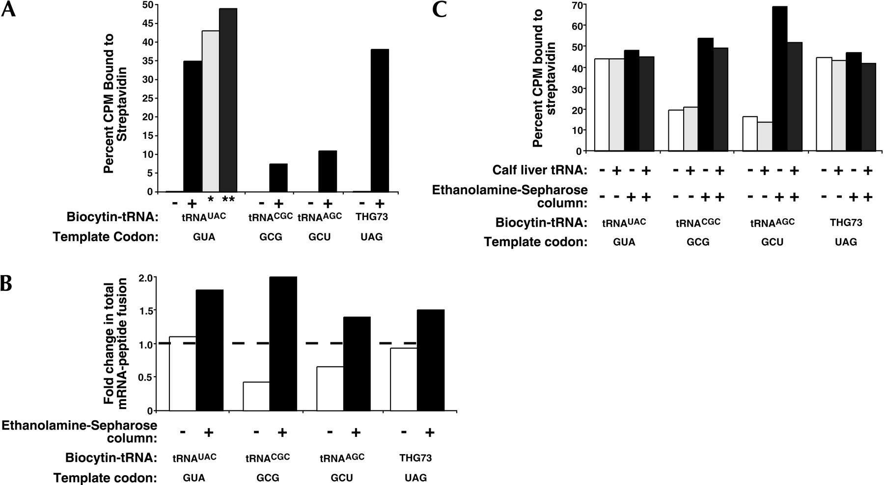
(A) Sense and nonsense suppression comparisons were performed in translation reactions of commercial rabbit reticulocyte lysate containing ~30 pmole of each template with fixed codons as indicated. The percent of [35S]Met-labeled mRNA–peptide fusions bound to immobilized streptavidin with or without translation reactions containing 2.0 μg of the indicated biocytin-acylated tRNA are shown. The binding results in which the translation lysate was bound to ethanolamine-Sepharose in batch mode to reduce endogenous tRNA concentrations before the suppression reaction is denoted by a single asterisk (*). The double asterisk (**) shows binding results from the same translation lysate supplemented with 0.5 μg calf liver tRNA. (B) The tRNA dependence of translation reactions as measured by the fold change in total mRNA–peptide fusion on the addition of 1.5 μg calf liver tRNA is used to assess the level of tRNA depletion of our in-house rabbit reticulocyte lysate preparation by column chromatography using ethanolamine-Sepharose. These values are calculated by dividing total [35S]-labeled mRNA–peptide fusions translated with supplemented calf liver tRNA by those fusions translated without supplementation. The broken line at 1.0 indicates no change in total mRNA–peptide fusion formation on the addition of calf liver tRNA. (C) The mRNA–peptide fusions described in B were then bound to streptavidin-agarose to determine the level of biocytin incorporation at the indicated template codons and expressed as the percent of [35S]Met-labeled mRNA–peptide fusions bound to immobilized streptavidin.




