


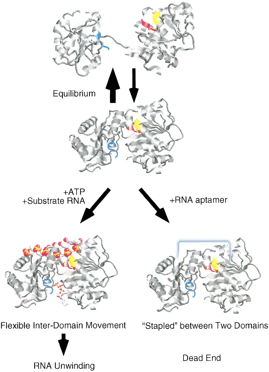
The schematic model for the mechanism of eIF4A inhibition by the RNA aptamer. We propose that eIF4A is in the equilibrium state between dumbbell and compact structures in solution, and that high-affinity RNA and ATP recognize the compact form and shift the equilibrium toward the latter. Whereas the ATP-bound state allows a flexible inter-domain movement between the amino- and carboxy-terminal domains necessary for the helicase action, the aptamer-bound state staples two domains and inhibits the conformational change necessary for ATP hydrolysis and the helicase action. Structures of full-length eIF4A are drawn in ribbon diagrams using the relevant coordinates of eIF4A (full-length, PDB ID code 1FUU; amino-terminal, ID code 1QDE; carboxy-terminal, ID code 1FUK) and RasMac Molecular Graphics. Motif Ia (PTRELA) is colored in yellow; motif II (DEAD), in red; and motif VI (HRIGRGGR), in cyan. In the bottom drawing, oligonucleotides and ATP are shown as space-filling model and ball-and-stick representation, respectively, according to the crystal structure of HCV NS3 protein complexed with oligo(dU) (PDB ID code 1A1V) and Caruthers et al. (2000).




