


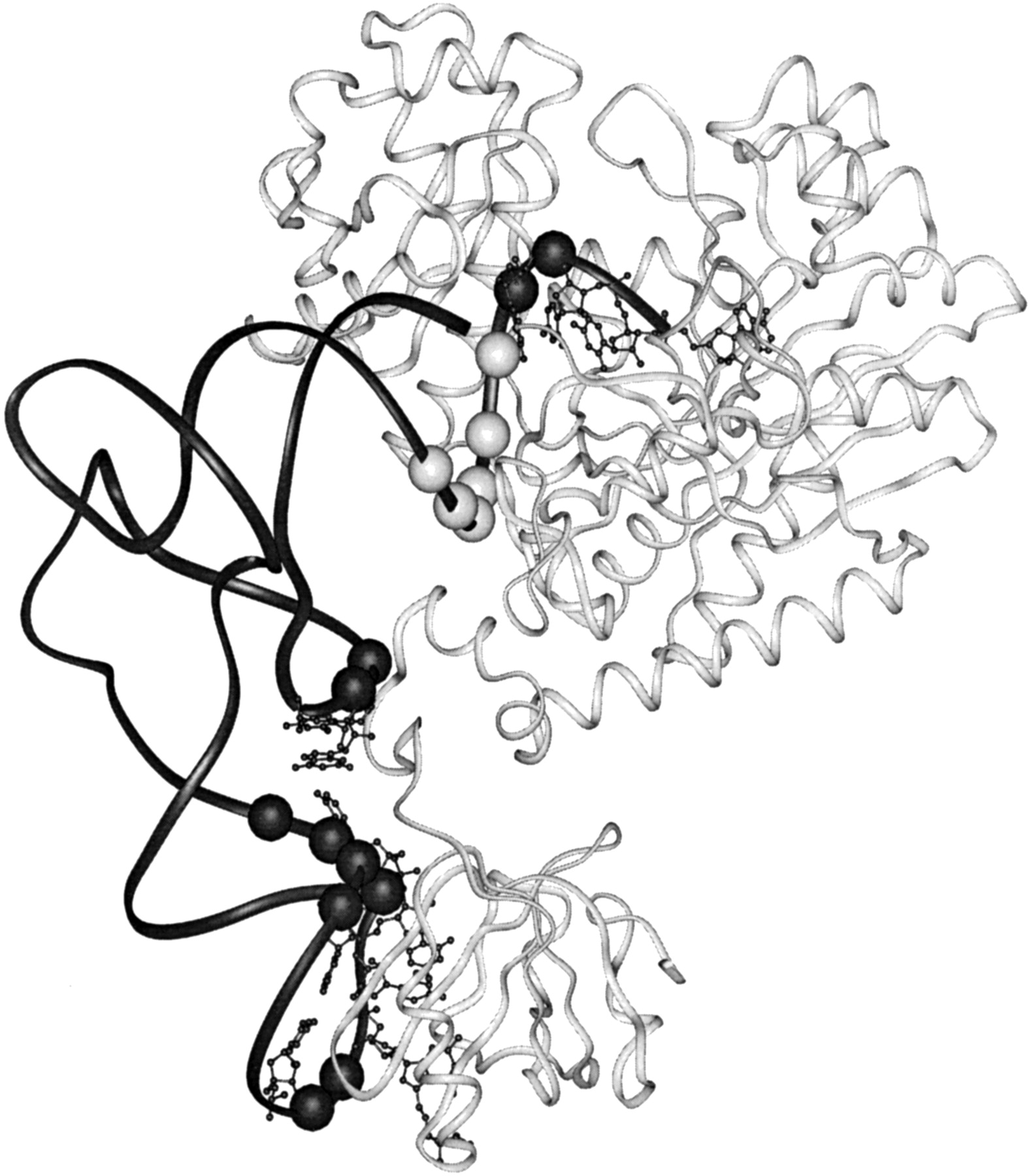
FIGURE 1.
Ribbon representation of one monomer of the dimeric E. coli AspRS-tRNAAsp complex showing the tRNA in black, with spheres representing phosphate sugar backbone residues that interact with AspRS; bases that interact with the protein are also shown. Note the contiguous series of six interacting phosphate sugar residues (light gray) in the tRNA acceptor stem (residues 67–71, upper right). The AspRS protein is shown in off-white (Eiler et al. 1999).




