


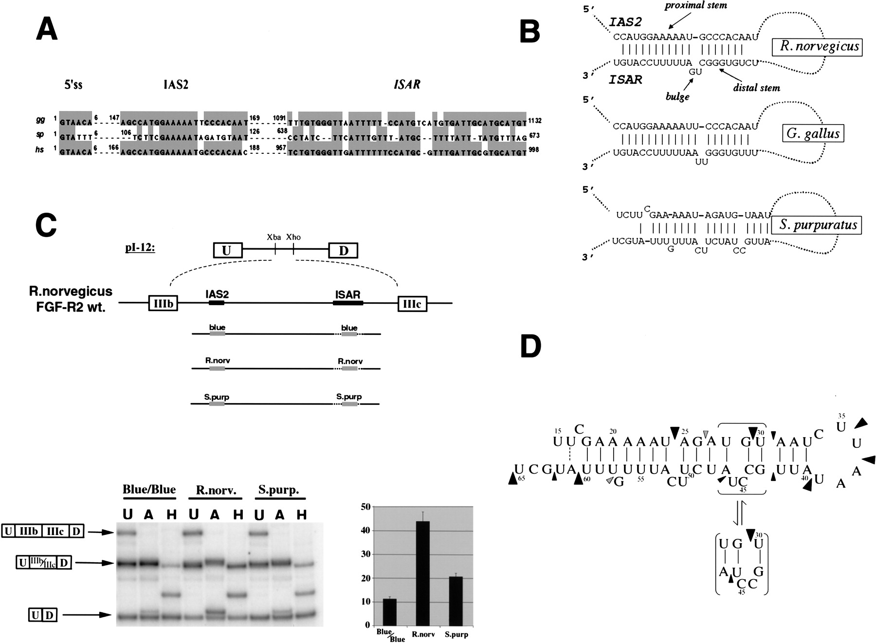
Conservation of IAS2–ISAR structure and function among FGF-R genes from sea urchins to humans. (A) A ClustalW alignment of intron sequences from H. sapiens, G. gallus and S. purpuratus downstream of exon IIIb revealed the conservation of several cis-acting elements. The 5′ splice site that borders exon IIIb and the conserved IAS2 and ISAR elements are shown for hsFGF-R2, ggFGF-R2, and spFGF-R genes. The position of the regulatory elements relative to the 5′ splice site (+1) is also indicated in each case. (B) Remarkable conservation of structure is apparent when comparing the potential for IAS2 and ISAR to form a secondary structure composed of two stems separated by a bulge (proximal and distal stems are indicated). (C) The spIAS2 and spISAR stem can partially substitute for the mammalian stem in an alternative splicing functional assay in rat DT3 cells (Carstens et al. 1998). The percentage of exon IIIb (with standard deviation) included is shown for a rat minigene construct were the rnIAS2 and rnISAR sequences were replaced with pBluescript sequences (Blue/Blue) as a negative control or replaced with the stem-forming portions of IAS2 and ISAR from either R. norvegicus or S. purpuratus. (D) An 89-nucleotide RNA, which included the spIAS2 and spISAR sequences separated by six nucleotides was synthesized by T7 RNA polymerase (see Materials and Methods). The structure of this RNA was probed with RNase A and RNase T1. Strong RNase A cleavage sites are indicated by large black arrowheads and weak sites are indicated by small black arrowheads. Weak RNase T1 cleavage sites are indicated by small gray arrowheads. The cleavage pattern shown is consistent with the specificity of the nucleases and the structure shown except for the RNase A cleavage 5′ of U30.




