


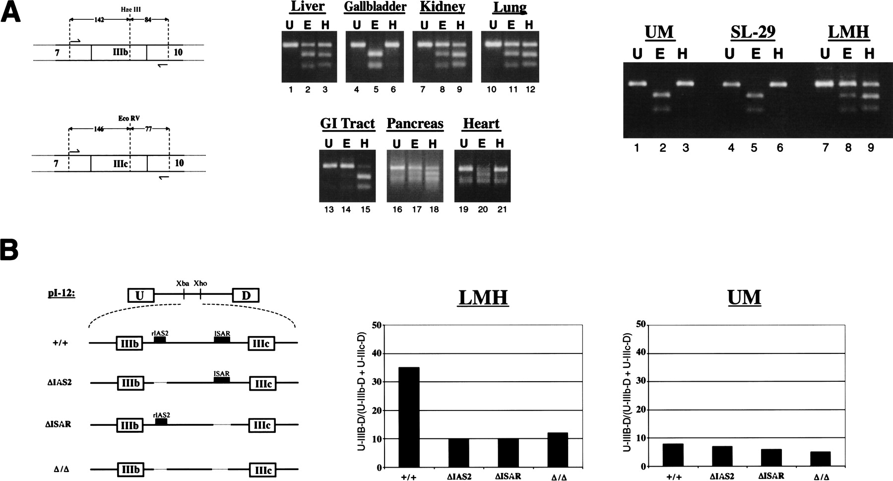
Chicken FGF-R2 transcripts are differentially spliced to IIIb or IIIc isoforms. (A) A schematic of the RT-PCR assay used to detect FGF-R2 transcripts and to distinguish between inclusion of IIIb and IIIc. Products of RT-PCR were separated on agarose gels and visualized by EtBr staining. RT-PCR products (U) were incubated with either HaeIII (H), which cleaves within exon IIIb, or EcoRV (E), which cleaves within exon IIIc. The middle panel shows RT-PCR analysis for chicken organs and right panel shows RT-PCR analysis for cell lines. Cell lines UM and SL-29 have a fibroblast morphology and cell line LMH has an epithelial morphology. (B) Schematic of four minigenes spanning the IgIII region of the rat FGF-R2 gene indicates the presence or absence of the IAS2 and ISAR (left panel). The level of exon IIIb inclusion from these minigenes transfected into LMH or UM cells is shown in the middle and right panels.




