


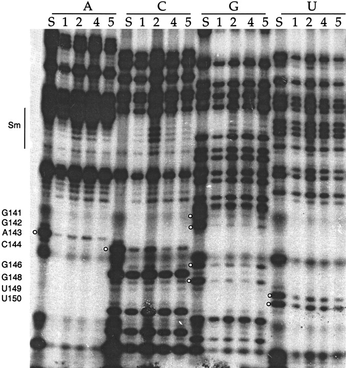
Iodine cleavage of RNAs containing phosphorothioates of A, C, G, or U shows interference at numerous positions near the 3′ end of U1. Numbers below each nucleotide indicate the complex (as in Fig. 2) from which the RNA is extracted. White balls indicate the stronger sites of interference in stem loop IV (see Fig. 1 for κ values). The numbers on the side represent nucleotide position. The Sm binding site sequence (nucleotides 125–133) is also indicated. S lanes are sample control lanes demonstrating phosphorothioate incorporation. Note that although some lanes may appear more intense because more RNA was loaded in the lane, these loading differences are accounted for in the calculation of κ. Occasionally, a common degradation band is seen (such as at position 145), excluding this nucleotide from analysis in this data set.




