


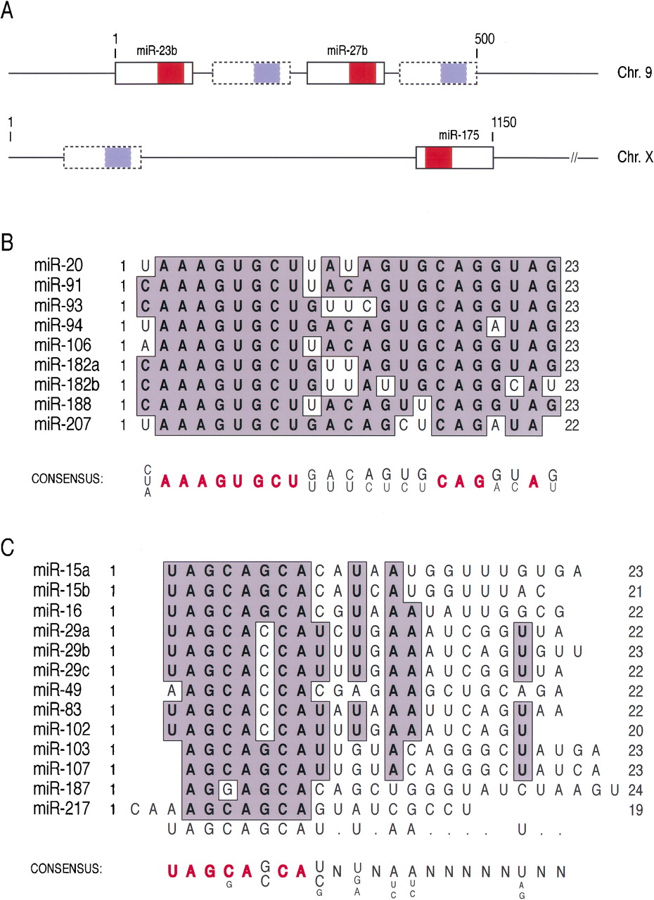
Novel miRNA clusters and families of miRNA paralogs associated with miRNPs in neuronal cells. (A) Novel miRNA clusters. The putative precursors of identified miRNAs are presented as solid boxes, and the mature miRNAs identified in the study are shown in red. Computer-predicted putative miRNA precursors are presented as dashed boxes, and the predicted mature miRNAs are shown in light blue. The chromosomal localization in the human genome is indicated on the right. The size of the region containing the miRNAs is indicated at the top of each cluster. (B) miRNPs contain a conserved miRNA family in neuronal cells. miRNA family members associated with miRNPs in neuronal cells are aligned. The names of miRNAs are indicated on the left. miRNA length is shown at the end of each miRNA. Conserved residues are in bold letters and gray shaded areas. Consensus is shown at the bottom: Nucleotides shown in red were invariable. (C) miRNPs contain a class of miRNAs with homologous 5′ ends in neuronal cells. miRNA class members with homologous 5′ ends are aligned. The names of miRNAs are indicated on the left. miRNA length is shown at the end of each miRNA. Conserved residues are in bold letters and gray shaded areas. Consensus is shown at the bottom: Nucleotides shown in red were invariable.




