


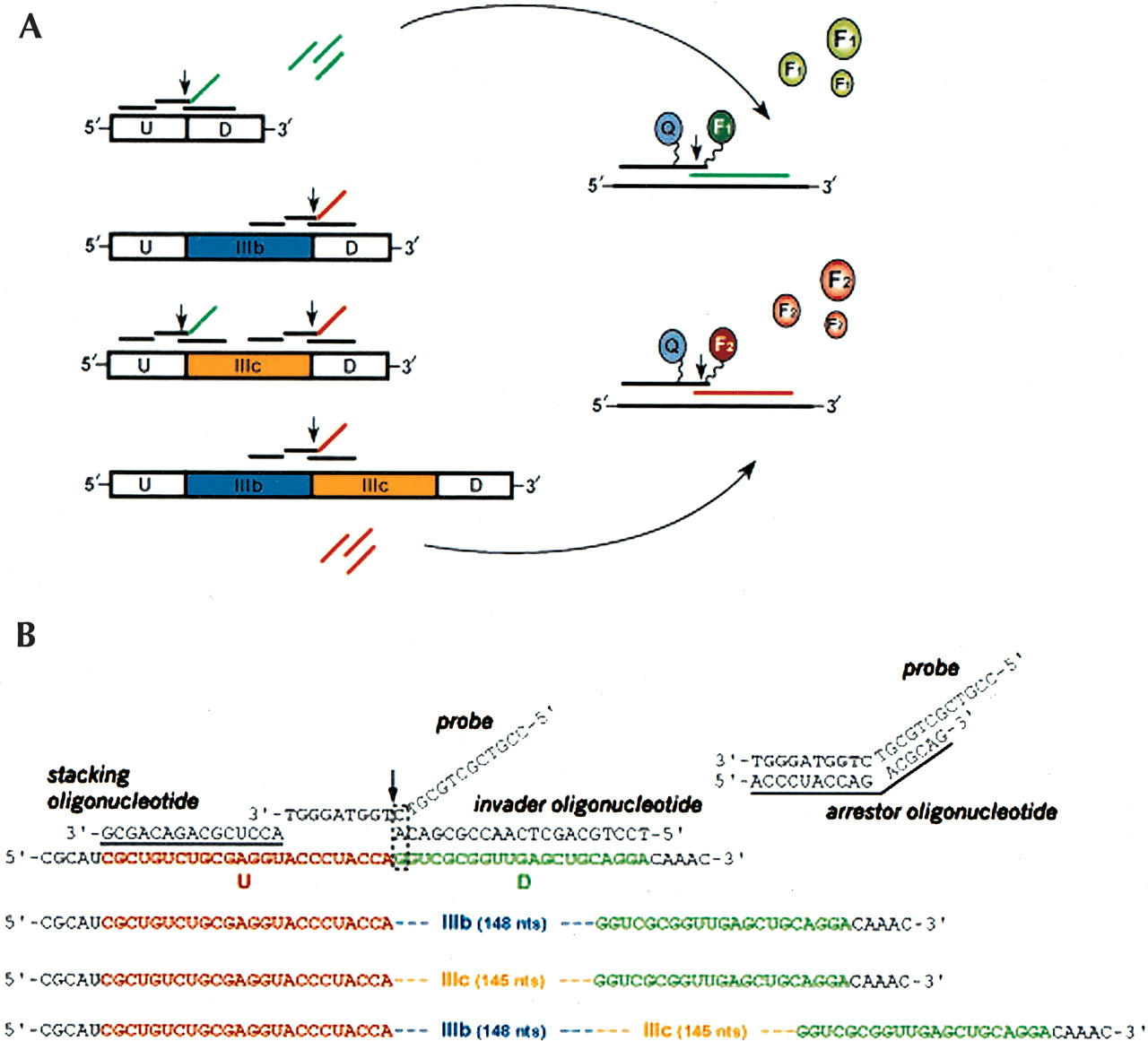
Schematics of the Invader RNA assay probe sets and the FGFR2 RNA variants derived from the alternative splicing of exons IIIb and IIIc. (A) Splice variants generated from the minigene constructs used in this report contain U and D exons, which are derived from Adenovirus2 L1 and L2 exons. Variants, depicted in the 5′–3′ orientation, can also contain IIIb and/or IIIc exons, which are derived from the rat FGFR2 gene (see Materials and Methods). Five Invader probe sets (see Table 1 for oligonucleotide sequences) were used to uniquely detect the splicing products. It should be noted that the IIIc-D probe set could also recognize this junction in the U-IIIb-IIIc-D RNA (not shown in the figure). Each probe set consists of, from left to right, a stacking oligonucleotide, the probe (with noncomplementary 5′ flap), and an Invader oligonucleotide (see B for more details). Two probe sets were run per Invader reaction using the biplex detection format (see Materials and Methods for the probe set combinations). Invader probe sets are shown in the 3′–5′ orientation as black lines above the splice variants except for the probe 5′ flaps, which are color-coded green or red to signify the fluorophore (F1 or F2) used for detection (right side of figure). Vertical arrows indicate Cleavase enzyme cleavage sites, which results in the release of multiple 5′ flaps (left side) or unquenched fluorophores (right side). Curved arrows indicate use of the cleaved 5′ flaps produced in the first reaction (left side) in the second reaction (right side). See text for further description of the Invader assay. (B) Detailed view of the U-D Invader probe set. The stacking oligonucleotide, probe, and Invader oligonucleotide are shown aligned on the U-D transcript from left to right. A dashed line box indicates the overlap structure and the vertical arrow indicates the probe cleavage site for the Cleavase enzyme. Transcript sequences for the IIIb, IIIc, and IIIb-IIIC splice variants are also shown to illustrate the inability of the probe and Invader oligonucleotide to form the overlap structure required for cleavage of the 5′ flap. A separate duplex structure between the probe and arrestor oligonucleotide, which is added in the secondary reaction to enhance signal generation, is depicted. Underlined sequences for the arrestor and stacking oligonucleotides indicate use of 2`-O-methylated nucleotides (see text). For further details see Eis et al. (2001) and de Arruda et al. (2002).




