


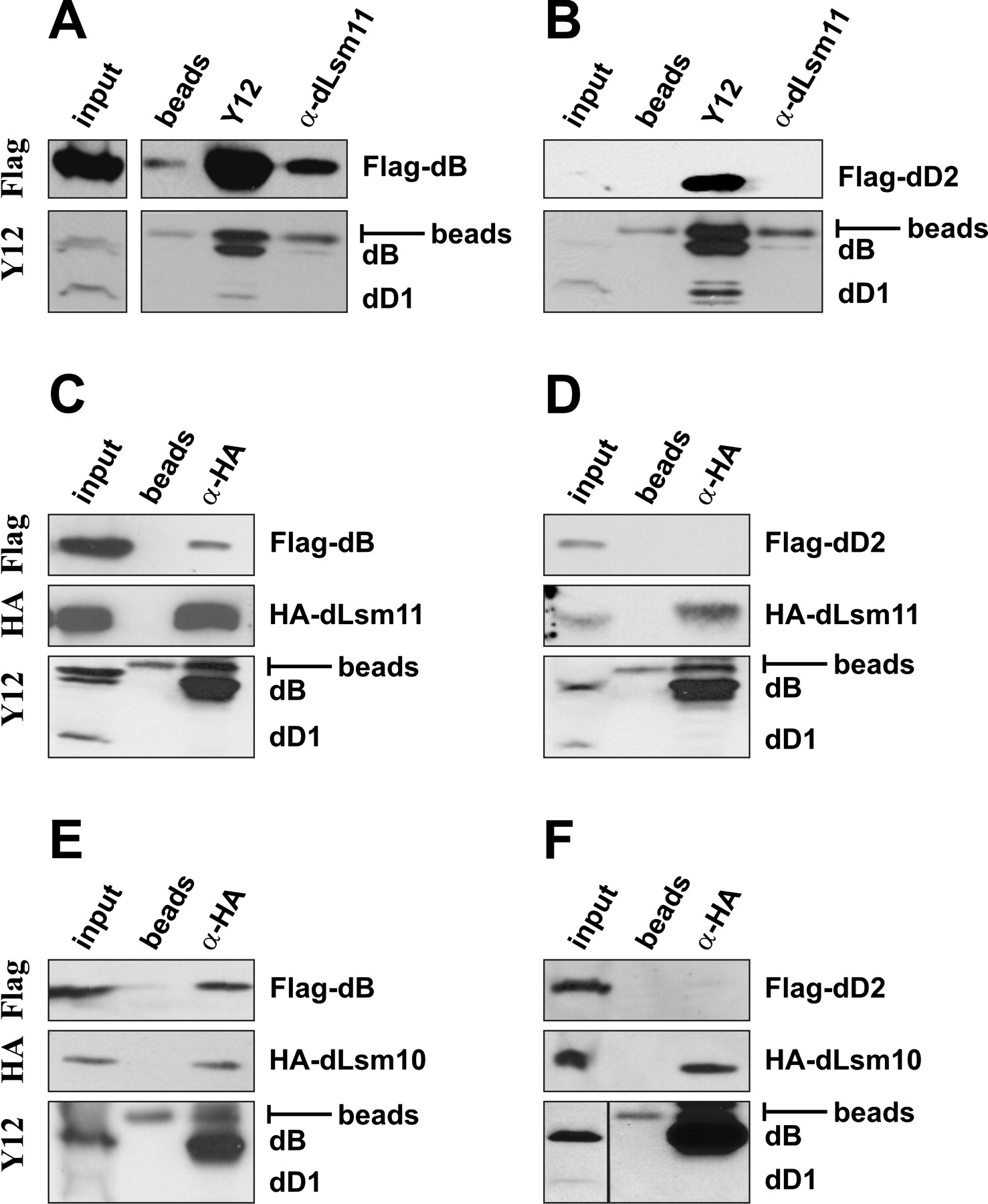
In vivo association of Drosophila Lsm10 and Lsm11 with Sm B, but not with Sm D1 and D2. Drosophila S2 cells were transiently transfected with expression plasmids encoding Flag-tagged Sm proteins dB (A, C, E) or dD2 (B, D, F) either alone (A, B) or in combination with HA-dLsm11 (C, D) or HA-dLsm10 (E, F). Whole-cell extracts were subjected to immunoprecipitation with the antibodies indicated above the lanes. The samples were subjected to SDS-PAGE, and Western blots were probed by using the antibodies indicated on the left of the figure. Input indicates sample of nuclear extract prior to immunoprecipitation; beads, control precipitation by protein G–Sepharose beads without antibody. A band corresponding to protein G released from the beads is visible in all the Y12 blots, just above the band specific for Sm dB. The black separation line in the bottom panel of F indicates that the input sample was run in a different position on this gel and had to be remounted.




