


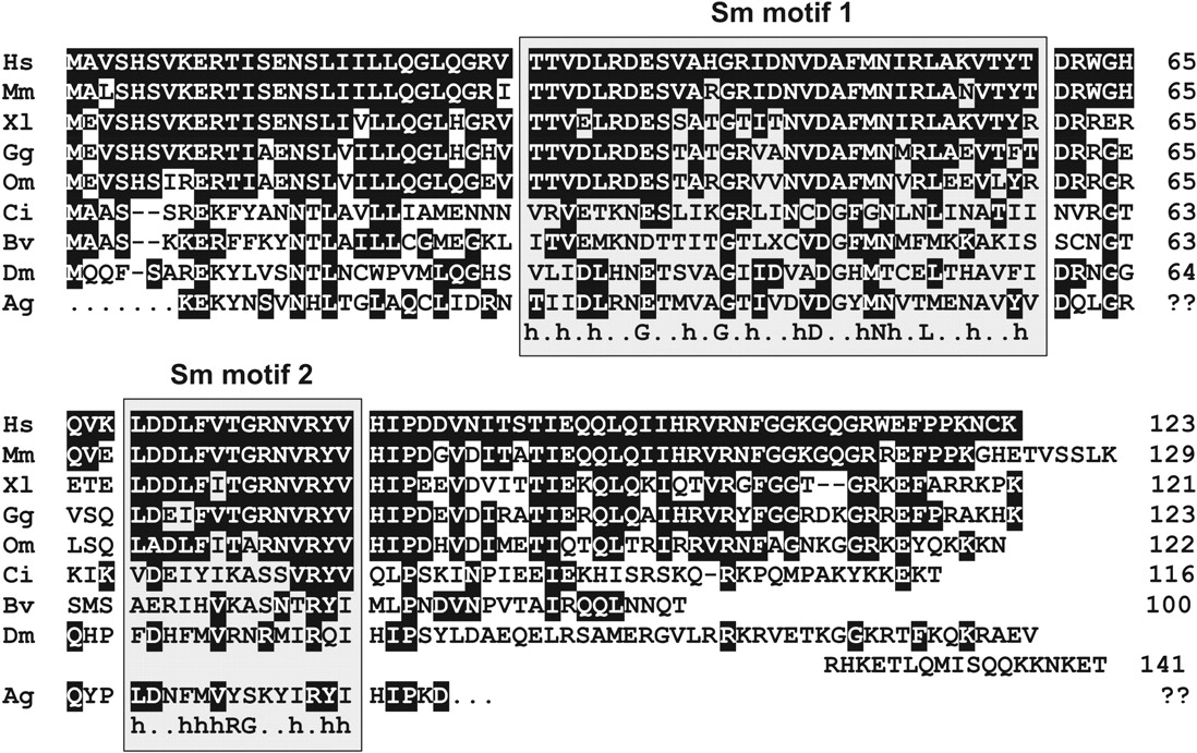
Sequence alignment of Lsm10 proteins. Additional sequences, accession numbers, and links are available at our online database (http://www.izb.unibe.ch/res/schuehome/schuemperli/Lsm10.html). The sequences are from the following species: Hs, Homo sapiens, human (Unigene Hs.3496); Mm, Mus musculus, house mouse (Unigene Mm.41865); Xl, Xenopus laevis (Unigene Xl.6554); Gg, Gallus gallus, chicken (ESTs BU211735, BU445159, BU461367); Om, Oncorhynchus mykiss, rainbow trout (EST CA368590); Ci, Ciona intestinalis, transparent sea squirt (ascidian tunicate; Unigene Cin.8743); Bv, Boltenia villosa (ascidian tunicate; protein AAM76159); Dm, Drosophila melanogaster (FlyBase CG12938); and Ag, Anopheles gambiae, African malaria mosquito (predicted partial ORF EAA01310). Residues identical to the human protein are shown in inverse print. The conserved Sm motifs 1 and 2 are indicated by shaded boxes. The consensus sequences were adapted from previous sources (Hermann et al. 1995; Achsel et al. 2001). h indicates hydrophobic amino acids.




