


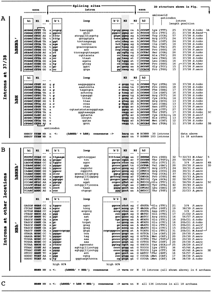
Intron–exon sequences in selected archaeal tDNAs sorted out as a function of the type of their splicing motifs. The details of the sequences of all introns presented in Figure 1 are given here. Additionally, all introns (any type) of S. tokodaii are illustrated. (A) Only 30 sequences of intron–exon junctions (out of a total of 103 sequences in the 800 tDNAs analyzed; see Supplementary Material) corresponding to intron located at position 37/38 are listed. The sequences shown extend from base 27 to base 43 of the anticodon arm. Uppercase letters correspond to exonic sequence and the lowercase letters to the intronic sequence. The bases in the H helix (under headings H1 and H2; gray background) are in bold; complementary bases in h and h′ helices (strands h1, h′1, h′2 and h2) are in bold italics. Mismatches in H helix are shown with inverted case: lowercase in H1 (exon) and uppercase in H2 (intron). Those that exhibit an intronic h′ helix of two or more base pairs are designated as canonical hBHBh′, and those with an intronic h′ helix shorter than 2 bp as relaxed hBH (see examples of 2D structures in Fig. 3). Notice that, for the hBHBh′ type, all elements of the splicing motif can be defined (h1, H1, B1, h′1, loop, h′2, H2, B2, and h2) whereas for the hBH type, only h1, h2, H1, H2, and B2 can be defined; the intronic h′1, h′2, and B1 elements cannot. At the bottom of A, is the consensus sequence of the splicing motif corresponding to the 30 sequences shown here or in all the 103 sequences (see Supplementary Material). Mismatched bases were not taken into account for consensus computation. For the degenerate alphabet used, see legend to Figure 1. (B) The 33 sequences of intron–exon junctions corresponding to introns located at positions other than 37/38 in the 800 tDNAs analyzed are listed as a function of the decreasing size of their h helix (exonic). The sequences shown extend from five bases upstream of the 5′ end of strand H1 down to five bases downstream from the 3′ base of the intron. The same lettering conventions as above are used. Those that exhibit an h helix of two or more base pairs are designated as canonical hBHBh′, and those with an exonic h helix shorter than 2 bp as relaxed HBh′. In this latter case, only H1, B1, h′1, h′2, and H2 are present and, at variance with the above hBH motif, no exonic h1, h2, and B2 elements can be defined. Vertical linking lines at right show the six cases of two introns in the same pre-tRNA. Numbering at the extreme right of the figure corresponds to the figure numbers in which the 2D structures are shown. At the bottom of B is the consensus sequence of the splicing motif corresponding to the 33 sequences listed in this panel. (C) generalized consensus from sequences shown in A and B and from all 136 introns in tDNAs of the genomes of the 18 archaeons analyzed. Mismatched bases in H helix were not taken into account for consensus computation.




