


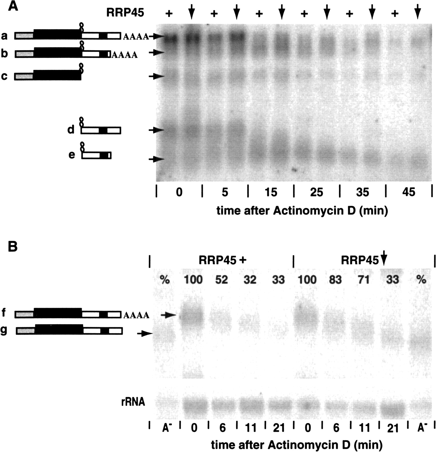
(A) The effects of RRP45 depletion on degradation of an RNA containing a strong secondary structure, G30C30, at the 3′-boundary of the CAT cassette. The experiment was performed as for Figure 3, and RNAs were identified using a probe hybridizing with the CAT gene (probe 1 in Fig. 2) and the EP1 3′-UTR (probe 2 in Fig. 2). The identities of the bands were determined using individual CAT and EP1 3′-UTR probes (not shown). As before, the plus sign (+) indicates that RRP45 was present at approximately normal levels (no tetracycline addition, no RNAi induction). The downward arrow indicates that the level of RRP45 was reduced (tetracycline present). (B) The effects of RRP45 depletion on degradation of the full-length CAT-EP1 RNA (analagous mRNA “a” in panel A, but without G30C30). The experiment was performed as for Figure 3, except that samples were run on a 2% agarose gel. Samples that were predigested with RNase H in the presence of oligo dT serve as markers for fully deadenylated transcripts (A−). The bands were detected with an RNA probe to the portion of the 3′-UTR that is absent in the shorter CAT-EP1 transcript (probe 3 in Fig. 2). The RNA probe cross-hybridized with ribosomal RNA, so we used this signal (lower panel) as a standard for phosphoImager quantitation, which is shown as percentage values on the lanes.




