


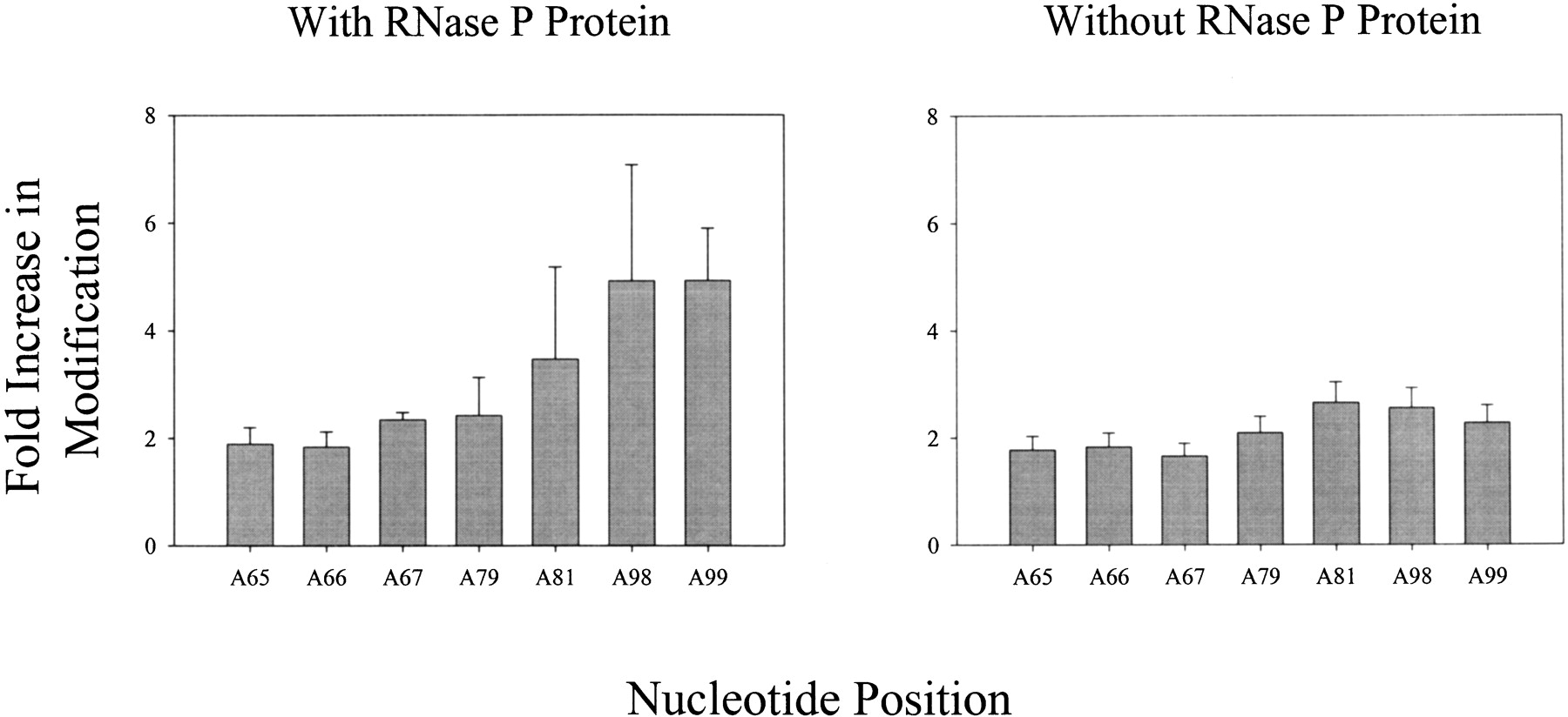
FIGURE 5.
Plots of the fold increase in DEPC modification at A residues 65–67, 79, 81, 98, and 99 in the presence (left) or absence (right) of RNase P protein when m(CAGCCUAC CCGG) is present.
(Downloading may take up to 30 seconds. If the slide opens in your browser, select File -> Save As to save it.)
Click on image to view larger version.
Plots of the fold increase in DEPC modification at A residues 65–67, 79, 81, 98, and 99 in the presence (left) or absence (right) of RNase P protein when m(CAGCCUAC CCGG) is present.
 CiteULike
 Delicious
 Digg
Digg Facebook
 Reddit
 Twitter
Molecular Cloning
The New Edition
Buy Now and Save 30%
Paperback only
(limited time offer)

|
Molecular Cloning: A Laboratory Manual (Fourth Edition) |
