


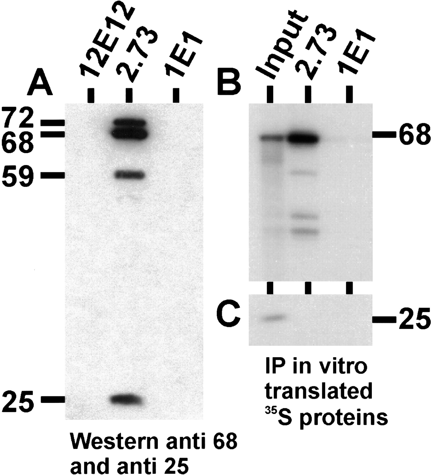
The anti-U1 70K antibody, mAb 2.73, immunoprecipitates CF Im from the light fractions. (A) The CF Im-rich light fractions were immunoprecipitated with mAb 2.73, mAb 12E12, and mAb 1E1. The immunoprecipitated proteins were separated by 10% SDS-PAGE and subjected to Western analysis probing with antibodies to both the 68-kD and 25-kD subunits of CF Im. The positions of the 72-, 68-, 59-, and 25-kD Cf Im subunits are shown. (B, C) 35S-met-labeled 68-kD (B) and 25-kD (C) subunits, prepared by in vitro transcription and translation, were immunoprecipitated with mAb 2.73 and mAb 1E1. The immunoprecipitated proteins were separated by 10% SDS-PAGE and autoradiographed. The input lane represents 1/10th of the amount of labeled proteins used for the immunoprecipitations.




