


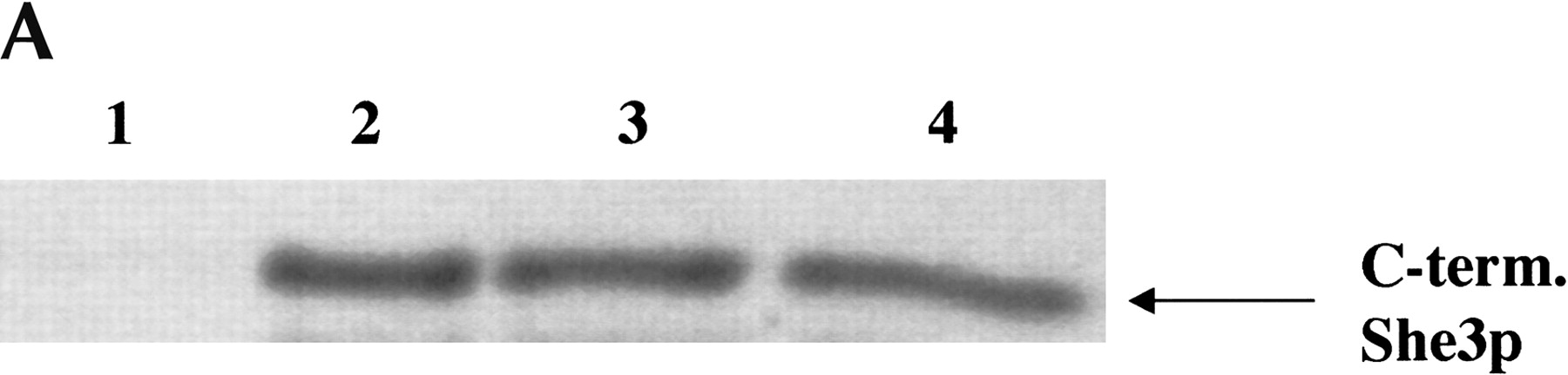
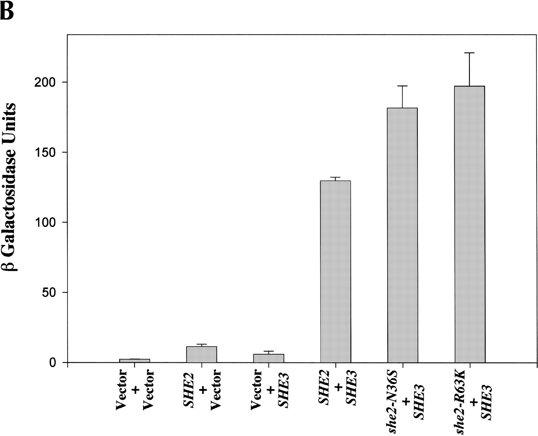
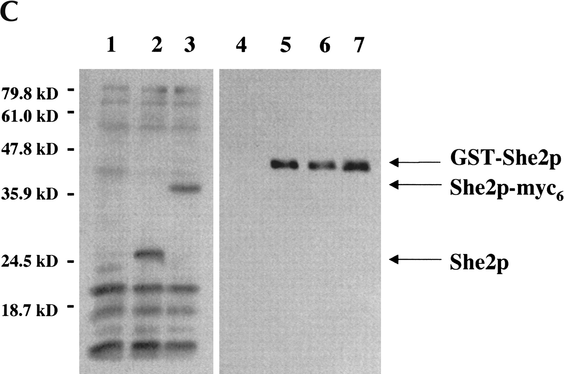
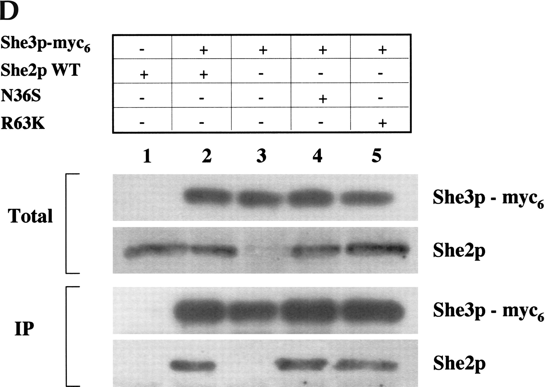
She2p mutants N36S and R36K are able to interact with She3p in vitro and in vivo. (A) In vitro GST pull-down assays were performed with 1 μg of (lane 1) GST, (lane 2) GST-She2p-wt, (lane 3) GST-She2p-N36S, or (lane 4) GST-She2p-R63K incubated with a C-terminal domain of She3p prepared by coupled in vitro transcription/translation. (B) She2p mutants N36S and R63K are able to interact with She3p by the two-hybrid assay. Strain PJ69-4a was transformed with the following combinations of plasmids: pGBDU-c1 (vector)/pACT2 (vector), pRL418 (pGBDU-c1-She2p-wt)/pACT2, pGBDU-c1/pEP13 (pACT2-She3p), pRL418/pEP13, pRL419 (pGBDU-c1-She2p-N36S)/pEP13, and pRL431 (pGBDU-c1-She2p-R63K)/pEP13. Transformants were grown in liquid culture and processed for β-galactosidase assays. (C) Characterization of the anti-She2p rabbit antiserum. Extracts were prepared from yeast strains YLM777 transformed with (lane 1) YCplac111 (vector), (lane 2) pRL453 (YCplac111-She2p), and (lane 3) pRL199 (YCplac111-She2p-myc6). Transformants of YLM777 were grown in synthetic media devoid of leucine. Lysates were prepared and used for Western blotting with anti-She2p rabbit antiserum. In addition, 5 ng of purified (lane 4) GST, (lane 5) GST-She2p-wt, (lane 6) GST-She2p-N36S, and (lane 7) GST-She2p-R63K were analyzed by Western blot with anti-She2p rabbit antiserum. The bands corresponding to She2p, She2-myc6, and GST-She2p are indicated by the arrows. (D) She2p mutants N36S and R63K associate with full-length endogenous She3p in vivo. Yeast strain YLM1320 was transformed with the following combinations of plasmids: (lane 1) YCplac22 (vector)/pRL453 (YCplac111-She2p-wt), (lane 2) pRL460 (YCplac22-She3p-myc6)/pRL453, (lane 3) pRL460/YCplac111 (vector), (lane 4) pRL460/pRL462 (YCplac111-She2p-N36S), or (lane 5) pRL460/pRL463 (YCplac111-She2p-R63K). Transformants were grown in synthetic media devoid of leucine as well as tryptophan, and lysates were prepared. An aliquot of the total lysate was analyzed by Western blot for She3p-myc6 and She2p (total). She3p-myc6 was immunoprecipitated from the lysate using anti-myc antibody, and the immunoprecipitates were analyzed by Western blot for the presence of She3p-myc6 and She2p (IP). She2p was detected in the immunoprecipitates using anti-She2 rabbit antiserum. Subsequently, the Western blot was stripped and reprobed with anti-myc to detect She3p-myc6.




