


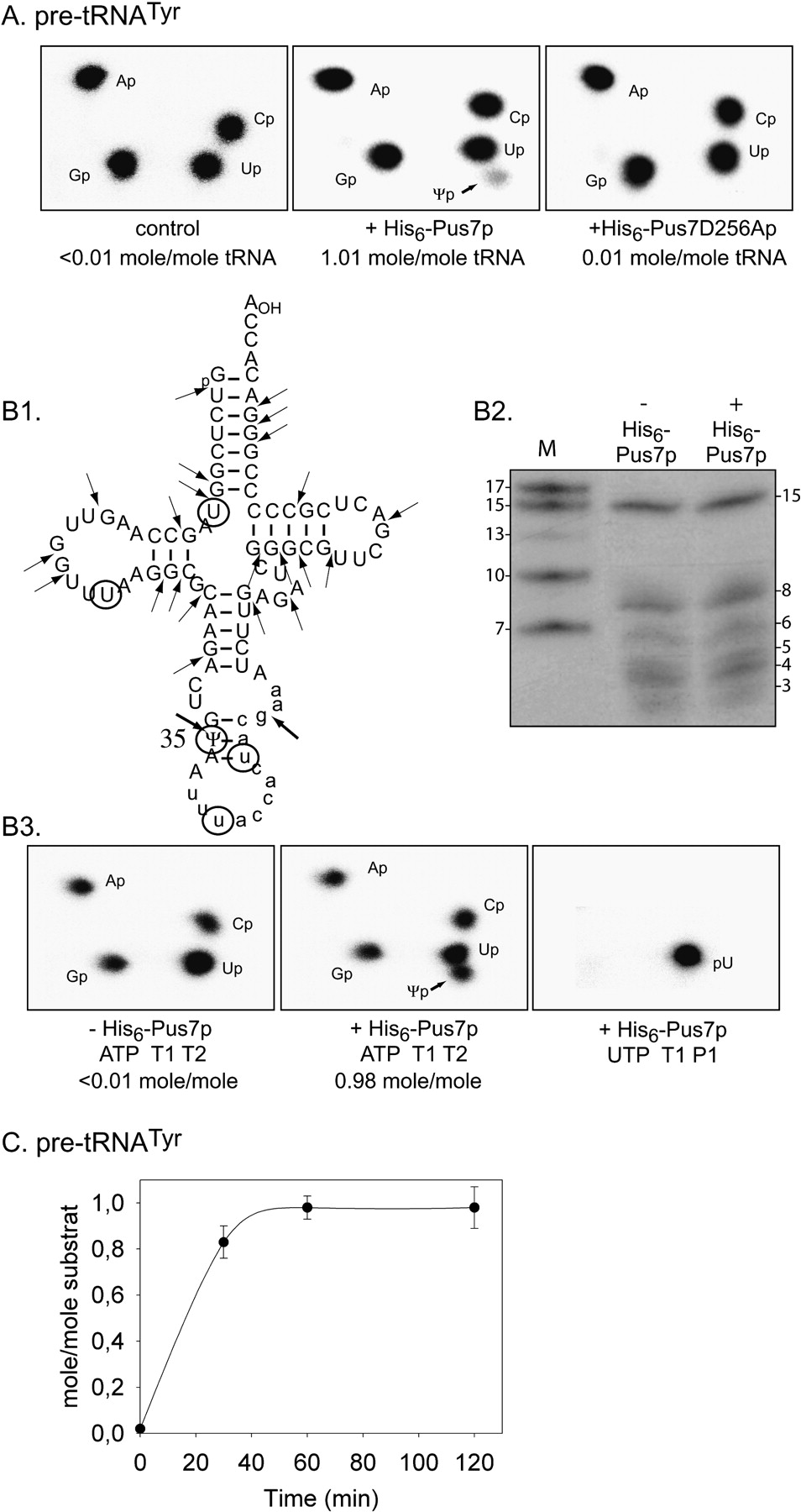
Recombinant His6–Pus7p catalyzes specifically the U to Ψ conversion at position 35 in yeast pre-tRNATyr. (A) Analysis of Ψ35 formation in pre-tRNATyr by the nearest neighbor approach. Yeast pre-tRNATyr uniformly labeled with [α-32P]ATP was incubated with the recombinant His6–Pus7p or His6–Pus7D256Ap variant in the conditions described in Materials and Methods. A control experiment was performed in the absence of recombinant protein. Modified and unmodified pre-tRNAsTyr were digested with T2 RNase and the 3′NMPs were fractionated by TLC. The autoradiograms of the TLC plates are shown. The molar ratio of Ψ residues formed in tRNAs, as deduced from quantification of the radioactivity of the spots, is given at the bottom of the panels. (B) Mapping of the Ψ residue formed in the pre-tRNATyr by recombinant His6–Pus7p enzyme. (B1) Cloverleaf representation of the pre-tRNATyr with indication of the T1 RNase cleavage sites. The U residues located at the 5′ position of an A residue are circled. (B2) Fractionation by gel electrophoresis of the T1 RNase products of the pre-tRNATyr labeled by [α-32P]ATP incorporation. Electrophoresis conditions are given in Materials and Methods. (B3) The 15-nt fragment obtained for a pre-tRNATyr incubated in the absence (− His6–Pus7p) or presence (+ His6–Pus7p) of the recombinant enzyme were digested with T2 RNase, and the resulting 3′NMPs were fractionated by 2D TLC. A similar experiment was performed with a pre-tRNATyr labeled by incorporation of [α-32P]UTP. In this case, the 15-nt T1 RNase digestion product was hydrolyzed with P1 nuclease and the resulting 5′NMPs were fractionated by 2D TLC. (C) Analysis of time-course formation of Ψ residue in the pre-tRNATyr upon incubation with the His6–Pus7p enzyme. pre-tRNATyr labeled by incorporation of [α-32P]ATP was incubated with His6–Pus7p enzyme in the conditions described in Materials and Methods. Aliquot fractions were collected at the indicated time after the beginning of the incubation. The pre-tRNATyr of each aliquot was digested with T2 RNase. The released 3′NMPs were fractionated by 2D TLC and the radioactivity of each 3′NMP was estimated by measurement with a PhosphorImager. (C) Represents the deduced ratio of Ψ residue moles formed per pre-tRNATyr moles as a function of the incubation times. Error bars were calculated as described in Materials and Methods.




