


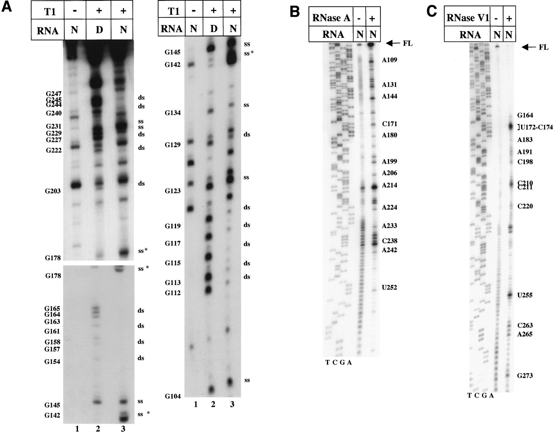
(A) RNase T1 probing of FMDV domain 3. [γ-32P]ATP-5′ end-labeled RNA corresponding to domain 3 was incubated with 0.03 U RNase T1 in native (N) or denaturing (D) conditions. Samples were analyzed on 6% acrylamide 7M urea gels run for different amount of time. Asterisks denote the residues with enhanced sensibility to T1; ss and ds stand for single-stranded or double-stranded, respectively. Bands with the same electrophoretic mobility than truncated fragments of the untreated transcript were not considered. (B) RNase A accessibility to domain 3 of the FMDV IRES. Domain 3 was incubated with diluted RNase A (5 × 10⁻⁶ mg/mL) to obtain a partial RNA digestion, and then subjected to primer extension analysis. (C) RNase V1 digestion of domain 3, analyzed by primer extension.




