


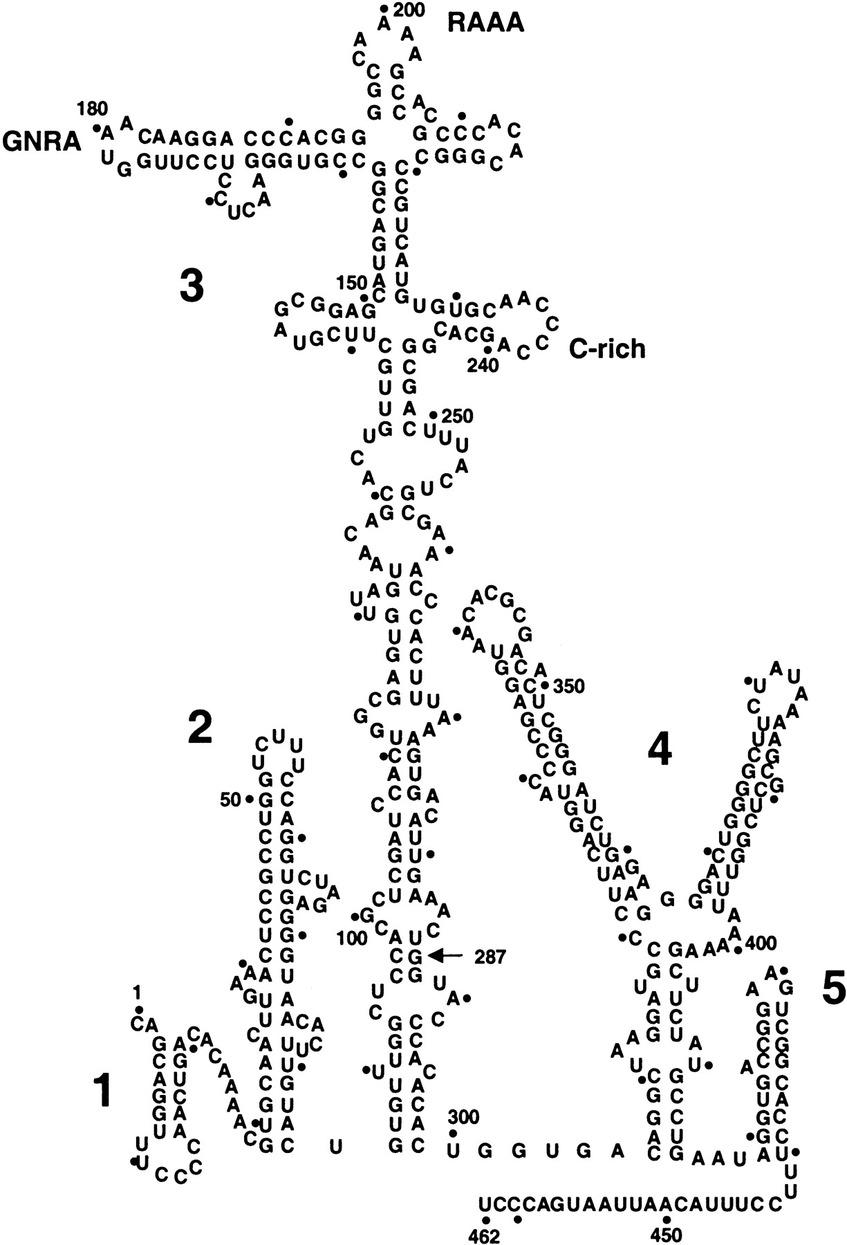
FIGURE 1.
Secondary structure of FMDV IRES. RNA structure was predicted using the Mfold program of GCG package, version 10. Domains are numbered 1 to 5, and span positions 1–85, 84–297, 306–415, and 416–462, respectively; a dot is used to mark positions each 10 nucleotides. The conserved GNRA, RAAA and C-rich motifs in domain 3 are highlighted. An arrow at position 287 points to the 3′ end of the antisense oligonucleotide used in primer extension assays.




