


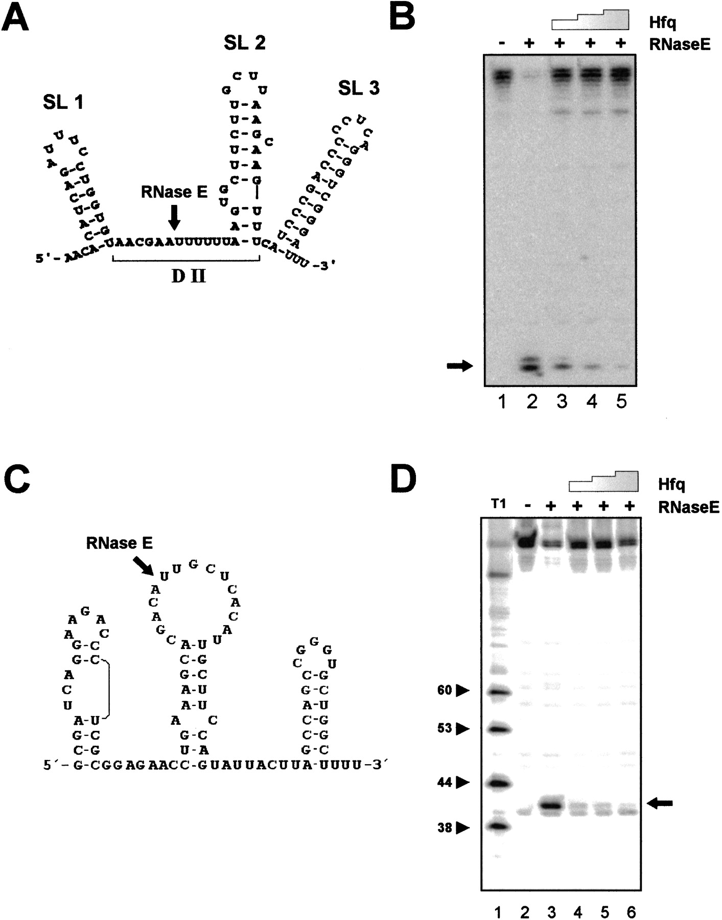
Hfq protects DsrA and RyhB sRNAs from RNase E cleavage. (A) The secondary structure of DsrA RNA was determined by Brescia et al. (2003). A ribo-oligonucleotide corresponding to Domain II (DII) inhibited binding of Hfq to DsrA (Brescia et al. 2003). The position of the RNase E cleavage site is indicated by an arrow. SL1, SL2, and SL3 denote the three stem–loop structures. (B) In vitro cleavage of DsrA RNA in the presence and absence of Hfq. [32P]-5′-end labeled DsrA was incubated in RNase E cleavage buffer at 37°C without (lane 1) or in the presence of a degradosome preparation (lanes 2–5). Hfq-hexamer was added in a fourfold (lane 3), 12-fold (lane 4), and 20-fold (lane 5) molar excess over DsrA prior to addition of the RNase E preparation. The RNase E cleavage reaction was carried out for 3 min at 37°C. (C) Secondary structure of RyhB RNA as revealed by Massé and Gottesman (2002) using bioinformatics. The RNase E cleavage site is indicated by an arrow. (D) In vitro cleavage of RyhB RNA in the presence and absence of Hfq. [32P]-5′-end labeled RyhB was incubated in RNase E cleavage buffer at 37°C without (lane 2) or in the presence of a degradosome preparation (lanes 3–6). Hfq-hexamer was added in a fourfold (lane 4), 12-fold (lane 5), and 20-fold (lane 6) molar excess over RyhB prior to addition of the RNase E preparation. The RNase E cleavage reaction was carried out for 3 min at 37°C. (Lane 1) RNase T1 ladder.




