


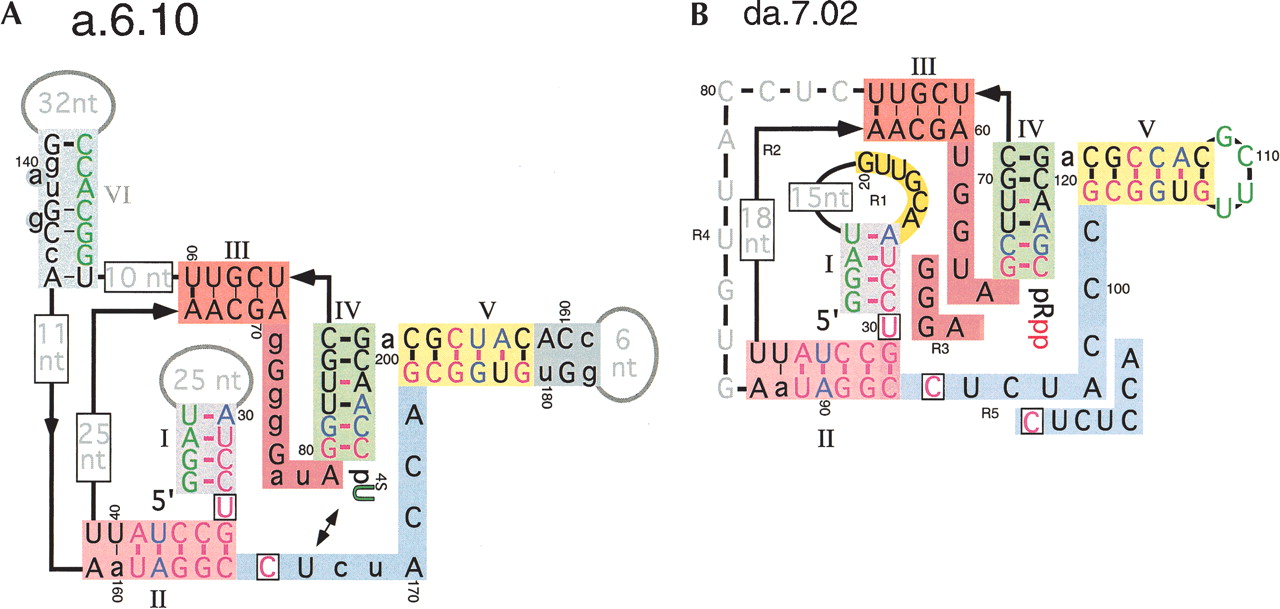
Inferred secondary structure of family A 4-thiouridine synthase ribozyme. (A) The structure of isolate a.6.10 obtained from the secondary structure determination selection is shown after having reacted with 4SUra to produce 4-thiouridine 5′ phosphate (p4SU). The ribozyme consists of six stems (I–VI), three terminal loops, and seven interhelical regions. U34 and C166 were absolutely conserved in every isolate sequenced (black boxed residues). Cross-linking the 4-thiouridine product of the ribozyme using UV light resulted in a efficient covalent linkage to U167 (arrows). Stems and conserved regions of sequences are denoted by colored boxes. Nucleotide colors: green, sequence fixed due to pool construction; magenta, no sequence variation observed (probability due to chance, P = 0.0012); blue, always covaries with partner (includes G-U wobble pairs). Uppercase black, ≤ 4/30 variations from consensus; lowercase black, > 4/30 variants from consensus. Residues showing poor sequence conservation are indicated in gray. Conserved potential for pairing is indicted as follows: thick purple bar, always base-paired (P = 3.6 × 10⁻⁵ if both residues can vary); thick black bar, 29/30 bp (P = 4.8 × 10⁻⁴); thin black bar, 26/30 or more bp (P<0.04). (B) The selection designed to minimize the ribozyme sequence in regions 1 to 5 (R1–R5) resulted in considerably shorter isolates, as represented by da.7.02. The consensus sequence for isolates resulting from round 7 of the selection differs from da.7.02 in R3 and R5 and is highlighted adjacent to the da.7.02 sequence. Conservation of sequence found in loop I is highlighted in yellow. The uppercase gray and boxed gray residues indicate variable sequence that could not be uniquely aligned, preventing the statistical analysis of mutational frequencies. The base-pairing information inferred from comparative analysis of the previous isolates (panel A) is shown within the helical regions of ribozyme da.7.02.




