


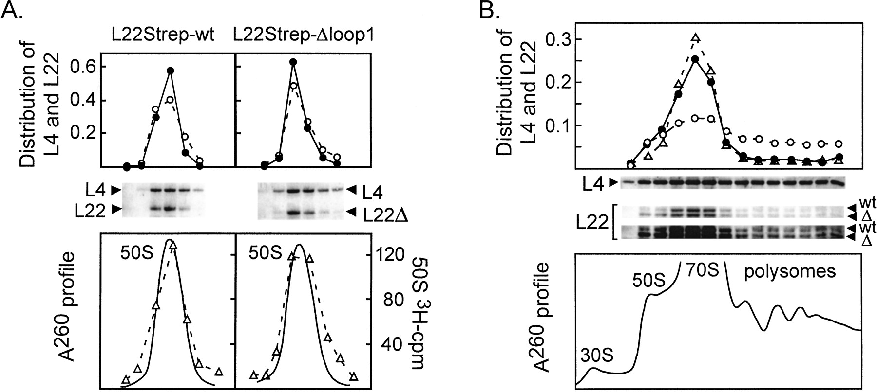
Sucrose gradient analysis of ribosomal particles containing wild-type or mutant L22 proteins. (A) Distribution of L22Strep and L22Strep-Δloop1 proteins in 50S ribosomes. Ribosomes enriched for 50S subunits were sedimented through a 10–30% sucrose gradient together with [3H]uridine-labeled purified 50S ribosomes prepared from a control culture. (Bottom) A260 profile together with a plot of the radioactivity in each fraction (indicated by open triangles and dashed lines). (Middle) Proteins from the collected fractions were fractionated on a 15% PAGE gel and subjected to Western analysis using anti-L4 and anti-Strep antibodies. (Top) For each fraction, the signal in chromosome-derived L4 or in the indicated plasmid-derived L22 protein was divided by the total signal for the protein in all of the fractions of the gradient. (○, broken line) Chromosome-derived L4; (•, solid line) plasmid-derived L22. (B) Sucrose gradient analysis of polysomes containing wild-type or mutant L22 proteins. (Bottom) A260 profiles. (Middle) Proteins from the collected fractions were separated on a 10% Tricine gel and subjected to Western analysis using anti-L4 and anti-Strep. Two different PhosphorImager exposures of the anti-Strep Western are shown. (wt) Wild-type L22-Strep; (Δ) L22Strep-Δloop1. (Top) For each fraction, the signal in chromosome-derived L4 or in the indicated plasmid-derived L22 protein was divided by the total signal for the protein in all of the fractions of the gradient. (○, broken line) Chromosome-derived L4; (▵, broken line) plasmid-derived wild-type L22; (•, solid line) plasmid-derived L22–Δloop1.




