


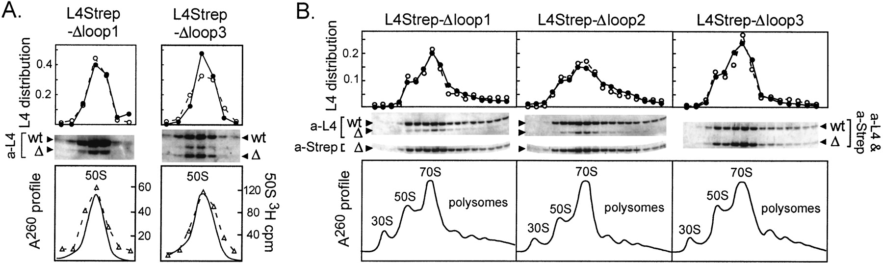
Sucrose gradient analysis of ribosomal particles containing L4 deletion proteins. (A) Distribution of L4 mutant proteins in 50S ribosomes. (Bottom) A260 profile together with a plot of the radioactivity in each fraction (indicated by open triangles and broken lines). (Middle) Western analysis using a 15% polyacrylamide gel and L4 antiserum. (Top) The signal in chromosome-derived L4 (wt) or in the indicated plasmid-derived extension-deletion protein (Δ) was calculated as described above. (○, broken line) Chromosome-derived L4; (•, solid line) plasmid-derived (mutant) L4. (B) Polysome gradient analysis. (Bottom) A260 profiles. (Middle) Western analysis of proteins from the collected fractions, probed with the indicated antibodies. (Top) For each fraction, the signal in chromosome-derived L4 (wt) or the indicated plasmid-derived extension-deletion protein (Δ) was divided by the total signal for the protein in all of the fractions of the gradient. (○, broken line) Chromosome-derived L4; (•, solid line) plasmid-derived (mutant) L4.




