


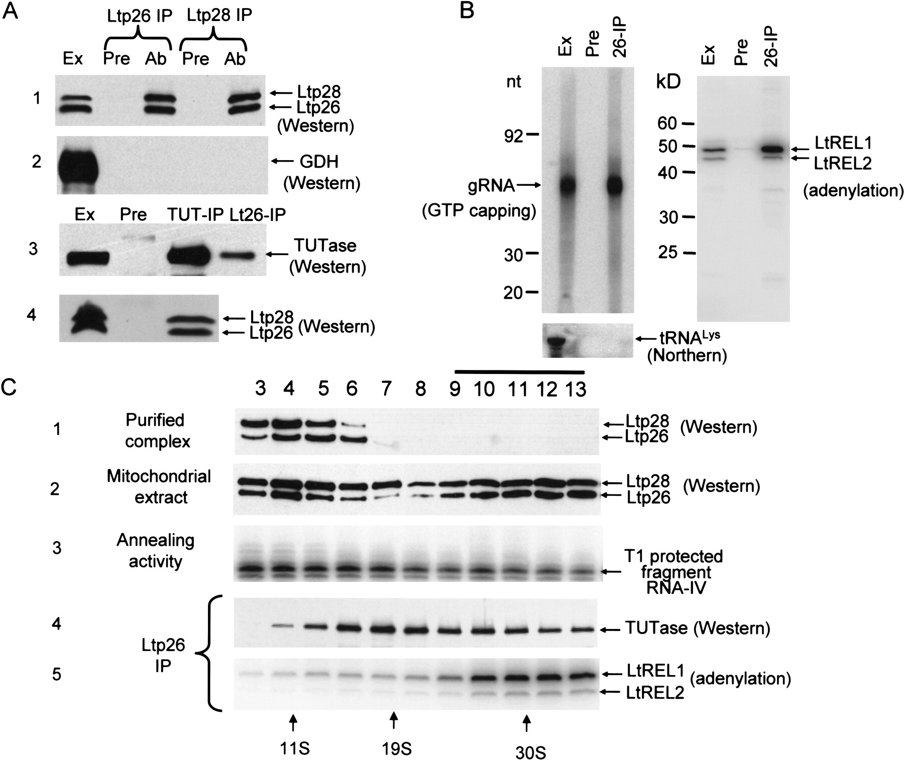
Association of Ltp26/Ltp28 complex with components of RNA editing machinery. (A) Co-IP of Ltp26/Ltp28 complex and 3′TUTase. IP from mitochondrial extract was performed using Sepharose G beads coated with affinity-purified mouse polyclonal antibodies against recombinant Ltp26, recombinant Ltp28, and L. tarentolae 3′ TUTase (Ltp26-IP, Ltp28-IP, and TUT-IP lanes). Immunodetection was performed with rabbit antibodies raised against purified Ltp26/Ltp28 complex, recombinant glutamate dehydrogenase (GDH) (Bringaud et al. 1997), and TUTase. Ext, mitochondrial extract; Pre, proteins bound to G-beads coated with preimmune serum; Ab, proteins bound to G-beads coated with specific antibodies. 10%–20% SDS gels blotted to nitrocellulose membranes and probed with respective antibodies. (B) Co-IP of Ltp26/Ltp28 complex, gRNA, and RNA ligase. IP was performed with preimmune (Pre) or anti-Ltp26 (IP) antibody. Left panel: Total RNA was isolated from mitochondrial extract and the IP material, labeled with [α32P]GTP in the presence of guanylyltransferase and separated on a 12% acrylamide/urea gel. Lower panel: The presence of tRNALys was assayed in the same material by Northern blotting with an oligonucleotide probe. Right panel: The same samples were incubated with [α32P]ATP to visualize the LtREL1 and LtREL2 proteins and analyzed on a 10%–20% SDS gel. (C) Association of Ltp26/Ltp28 complex with TUTase and with RNA ligase. Purified Ltp26/Ltp28 complex (10 μg; panel 1) and mitochondrial extract (250 μL, 10 mg/mL; panels 2,3) were fractionated on a 10%–30% glycerol gradient in the SW41 rotor for 20 h at 35,000 rpm. Panel 2, Western analysis of Ltp26 and Ltp28. Panel 3, T1 nuclease protection assay for RNA annealing activity. Panels 4 and 5, co-IP of TUTase and LtREL1 and LtREL2 by anti-Ltp26 antibody.




