


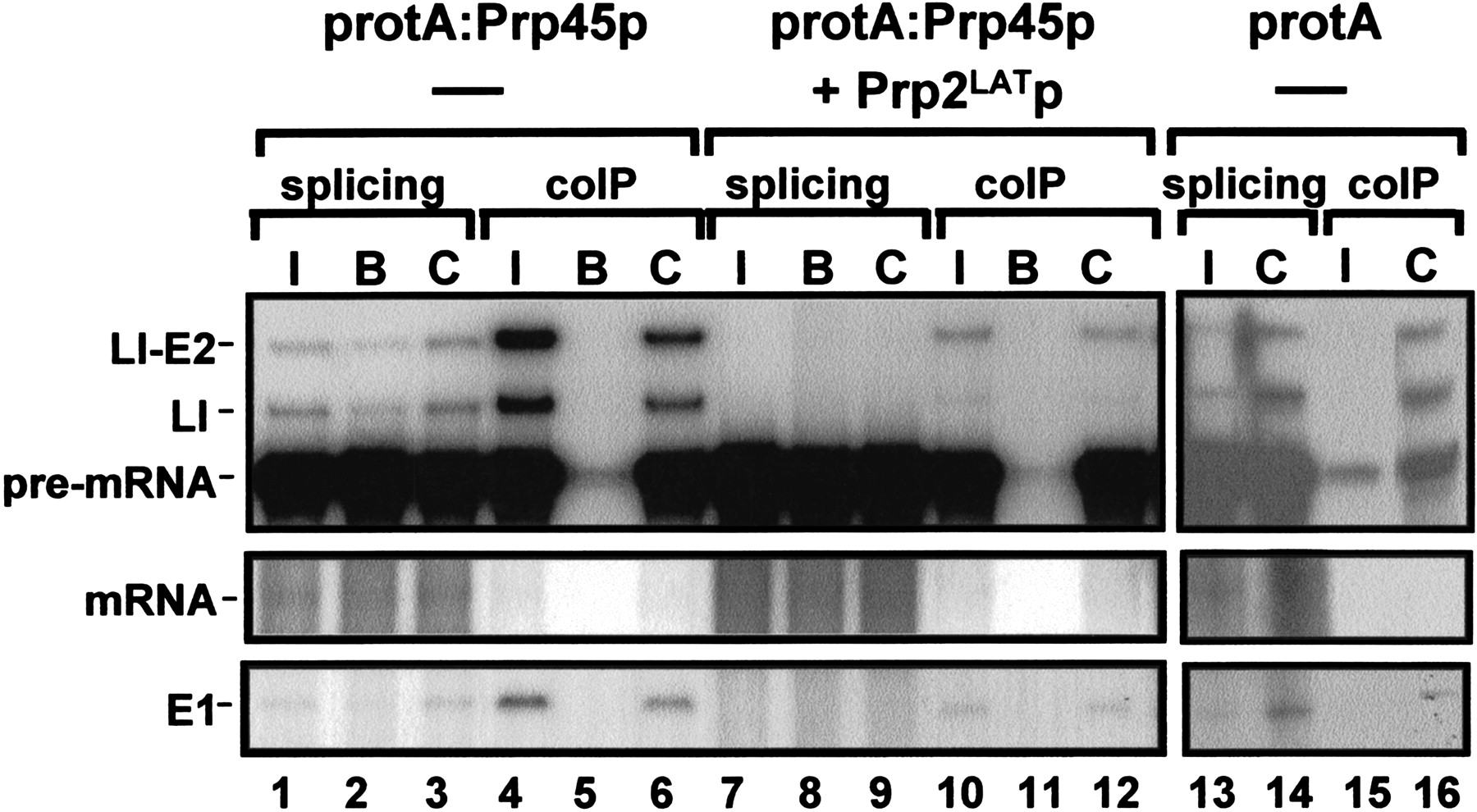
Coprecipitation of spliceosomes by Prp45p. Whole yeast cell extract (splicing extract) was prepared from cells of strain YMA45/2 grown in galactose-based medium, producing protA:Prp45p. As a control, extract was prepared from the parental wild-type strain carrying vector pNOPPATAIL, producing a double protein A epitope (protA). Splicing (50 μL total volume) was performed using 32P-labeled actin pre-mRNA. The reactions were stopped and 5 μL were removed as splicing controls (input). The remaining 45-μL samples were mixed with an equal volume of precipitation buffer containing either IgG-agarose beads (I), agarose beads without antibody (B) or protein A-Sepharose beads with prebound anti-Prp8p antibodies (C) and incubated at 4°C for 2 h. Beads were washed in buffer containing 150 mM NaCl, deproteinized, and the RNAs precipitated. The samples from the immunoprecipitations (coIP) as well as from the input samples (5 μL; splicing) were then resuspended in formamide loading buffer, resolved on a 6% (w/v) polyacrylamide gel, and labeled RNAs were visualized by autoradiography. In additional samples, recombinant dominant negative Prp2p was added to the extract prior to splicing (+ Prp2LATp) and the samples were treated as above. The positions of the RNA species are indicated. (lariat I-E2) Lariat intron-exon2; (LI) Lariat-intron; (E1) exon1.




