


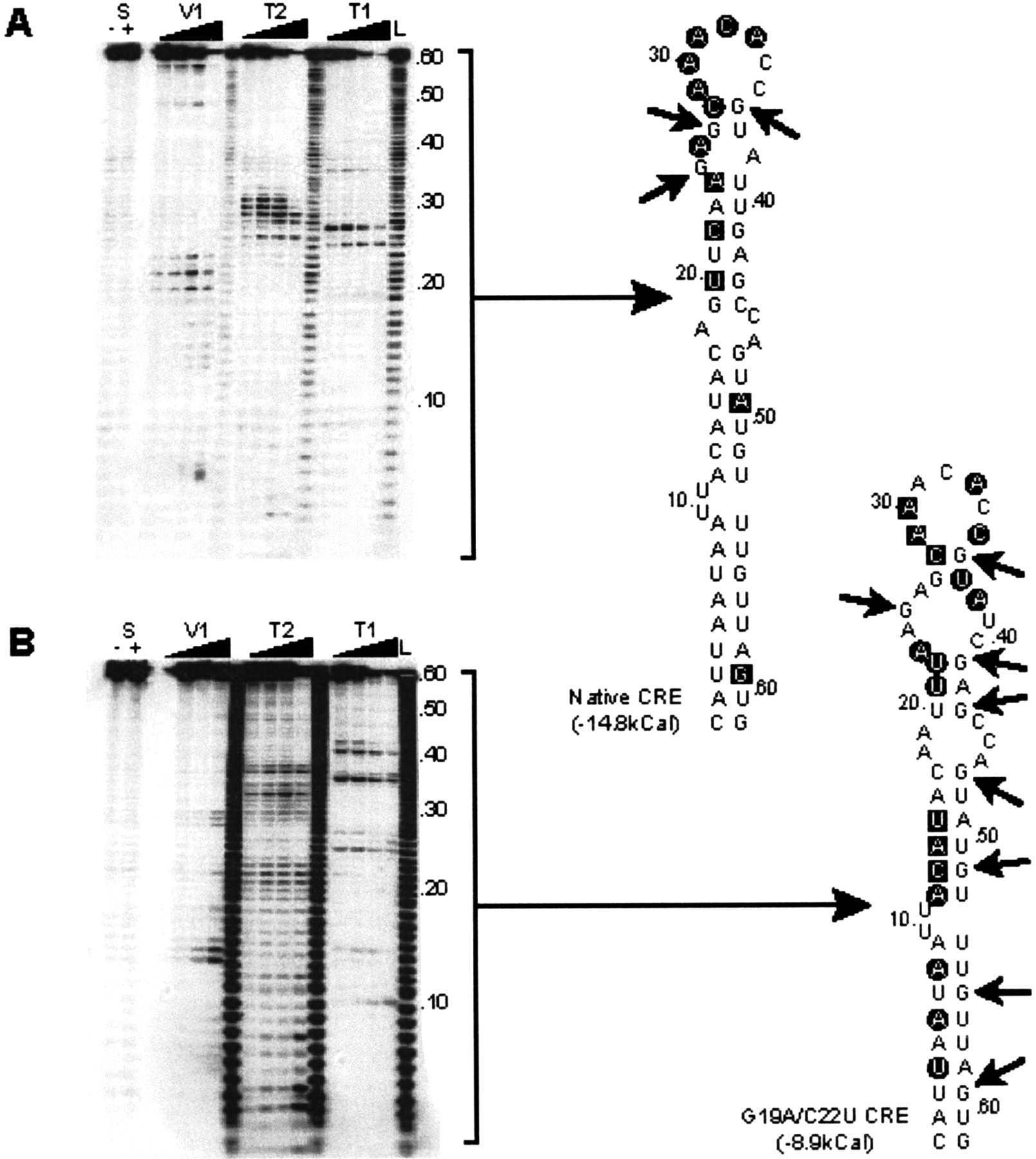
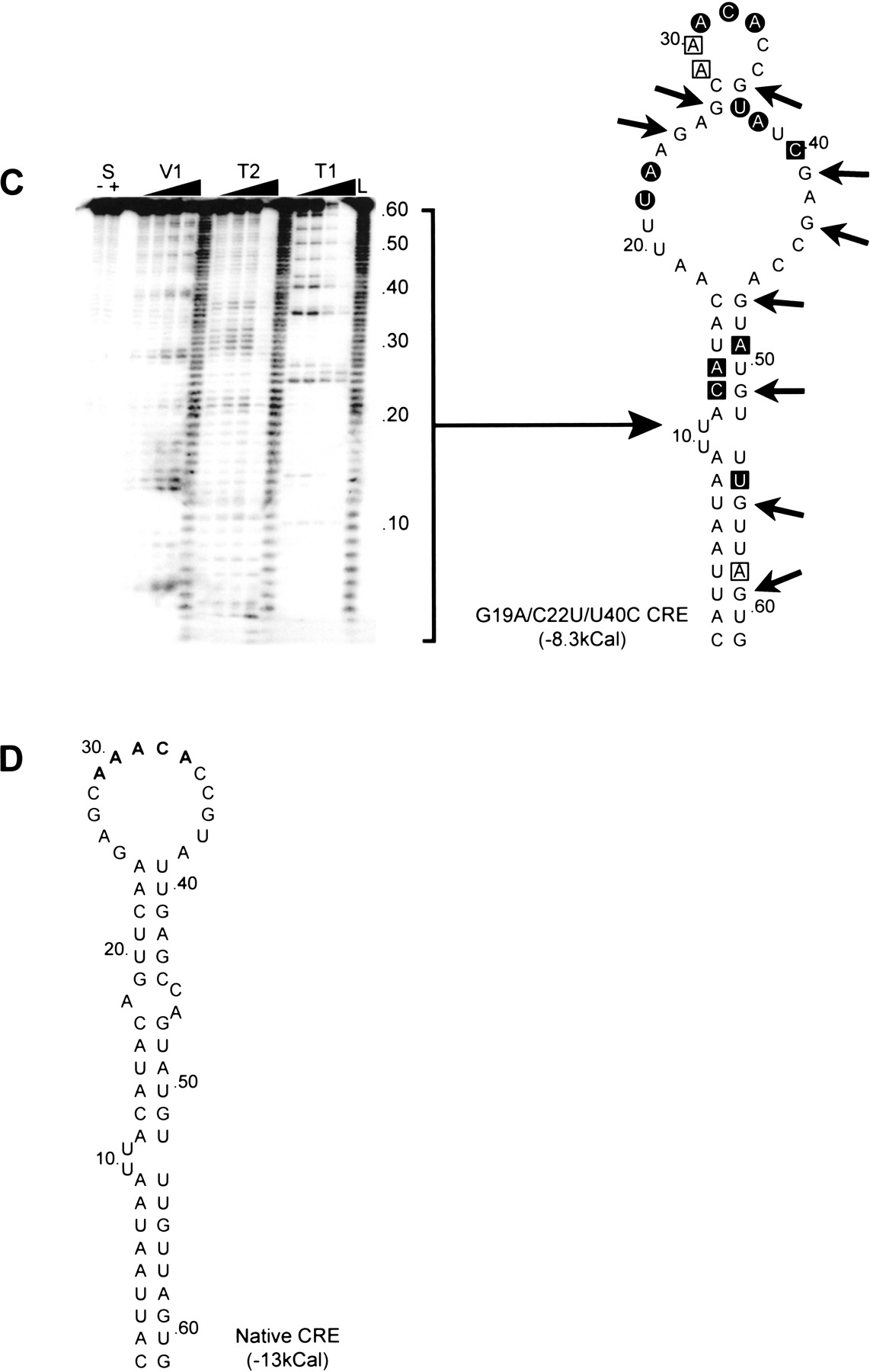
Structural mapping of the poliovirus CRE. (A) The autoradiograph of a representative mapping acrylamide gel is shown alongside a diagram of the ribonuclease cleavages indicated on the predicted structure of the CRE. T1, V1, and T2 cleavages are indicated with an arrow, black square, or black circle, respectively. The few positions cleaved with both V1 and T2 are indicated with an open square. Lane L is a hydroxidegenerated 1bp ladder produced from the same probe, lanes S are the probe alone incubated in the absence (−) or presence (+) of 100 mM sodium chloride (which is required for V1 ribonuclease activity). (A) The native poliovirus type 3 CRE. (B) The poliovirus type 3 CRE containing two substitutions in the Stem 3 region (see Fig. 1A) of G19A and C22U. (C) The poliovirus type 3 CRE containing three substitutions in the Stem 3 region of G19A, C22U, and U40C. (D) The proposed structure of the native poliovirus type 3 CRE derived from ribonuclease mapping studies.




