


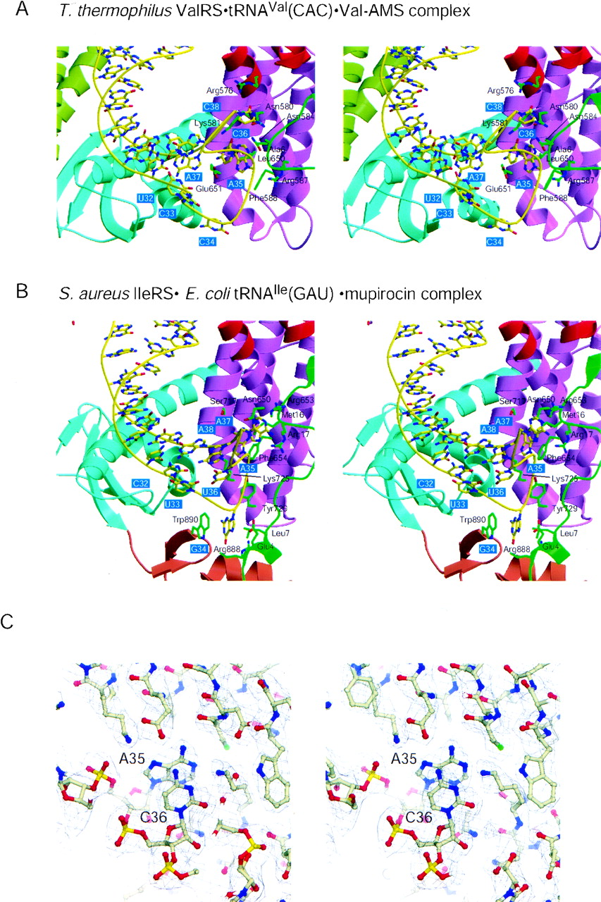
(A) Interactions between ValRS and the tRNAVal anticodon arm (stereoview). Each domain of ValRS is colored as in Figure 1. The trace of the C1′ atoms of tRNAVal is shown as a yellow tube. The bases of tRNAVal are shown as sticks, where the carbon atoms are yellow. The tRNA-interacting amino acid residues of ValRS are shown as sticks, where the carbon atoms are green. A35 and C36, which are the major identity elements of tRNAVal, are recognized by the α-helix bundle domain. (B) Interactions between IleRS and the tRNAIle anticodon arm (stereoview). The representation is as in Figure 3A. G34 is recognized by the C-terminal wobble-base-binding domain. (C) σa-weighted Fo-Fc omit map around A35 and C36 of tRNAVal, contoured at the 5.0σ level (stereoview). A35, C36, and the surrounding amino acid residues were omitted. The atomic model of the ValRS•tRNAVal•Val-AMS complex is shown as a ball-and-stick representation.




