


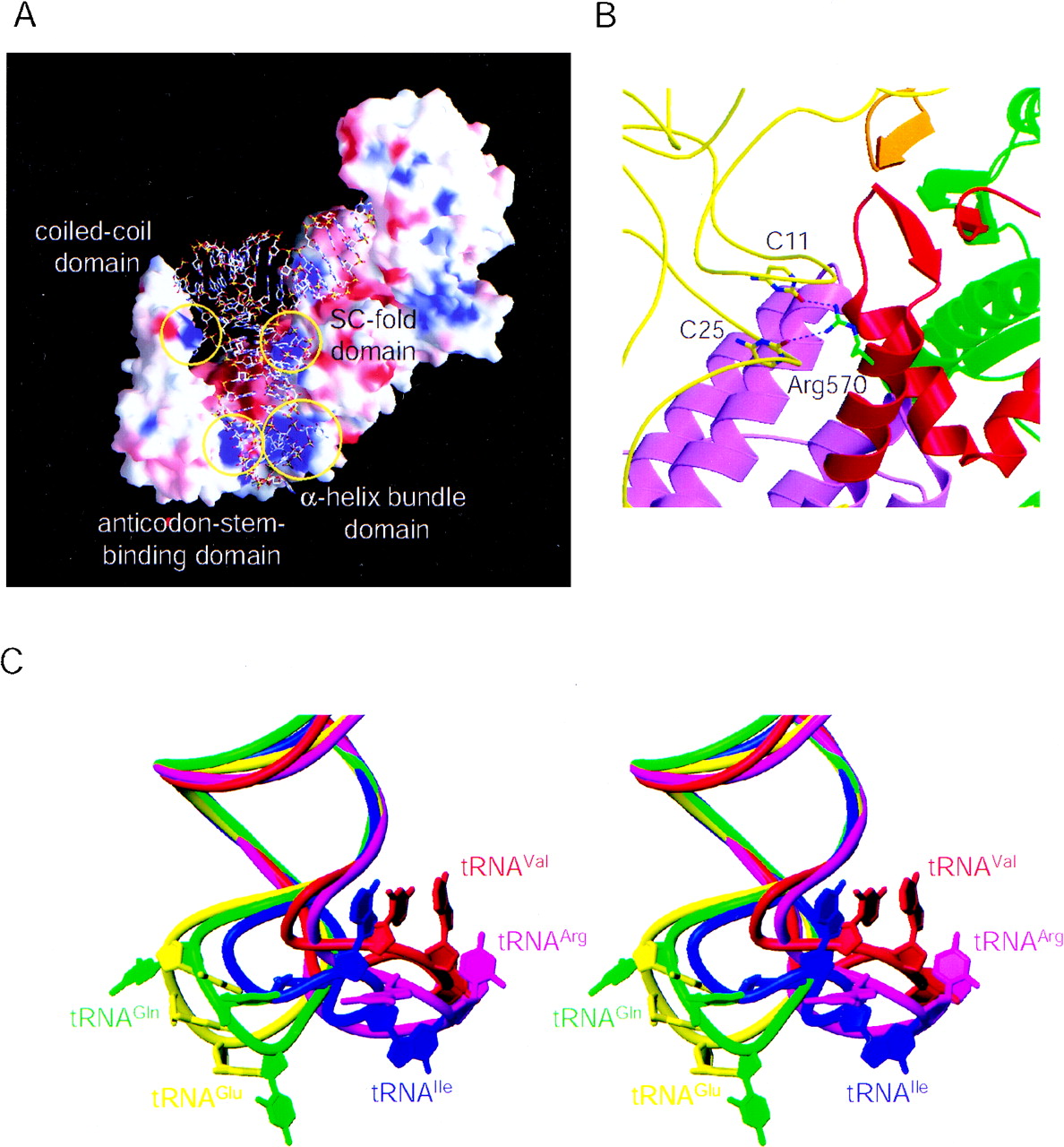
(A) Surface potential of the T. thermophilus ValRS. (B) Interactions of the ValRS SC-fold domain with the tRNAVal D stem. Each domain of ValRS is colored as in Figure 1. The trace of the phosphorus atoms of tRNAVal is shown as a yellow tube. C11 and C25 of tRNAVal are shown as sticks, where the carbon atoms are yellow. Arg570 of ValRS is shown as sticks, where the carbon atoms are green. (C) The anticodon-loop structures of tRNAs bound to the class Ia and Ib aaRSs (stereoview). The phosphorous atoms of the anticodon stems are superposed. This figure shows the clear difference in the anticodon-loop structures between the tRNAs bound to the class Ia and Ib aaRSs.




