


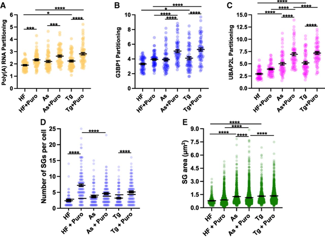
Comparative analysis of candidate stress granule properties in cells treated with halofuginone, arsenite, or thapsigargin in the presence or absence of puromycin. Partitioning coefficients of polyadenylated RNA (A), G3BP1 (B), or UBAP2L (C) from FISH/immunofluorescence images shown in Figure 4 (n = 60 cells). Cells were treated with halofuginone (2 μM, 4 h) with or without puromycin (10 μg/mL) added during the last 30 min or arsenite (25 μM, 1 h) or thapsigargin (0.04 μM, 1 h) with or without puromycin (10 μg/mL). Results are from three to four frames per replicate per condition from three independent experiments. (D) Number of stress granules per cell in cells treated as in A–C. Stress granules (UBAP2L staining) within 5 µm of the nuclear boundary were quantified. (E) Stress granule area (UBAP2L staining, within 5 µm of nuclear boundary) was determined. One-way ANOVA with Tukey HSD tests were done to assess significance with (*) P < 0.05, (***) P < 0.005, (****) P < 0.001.




