


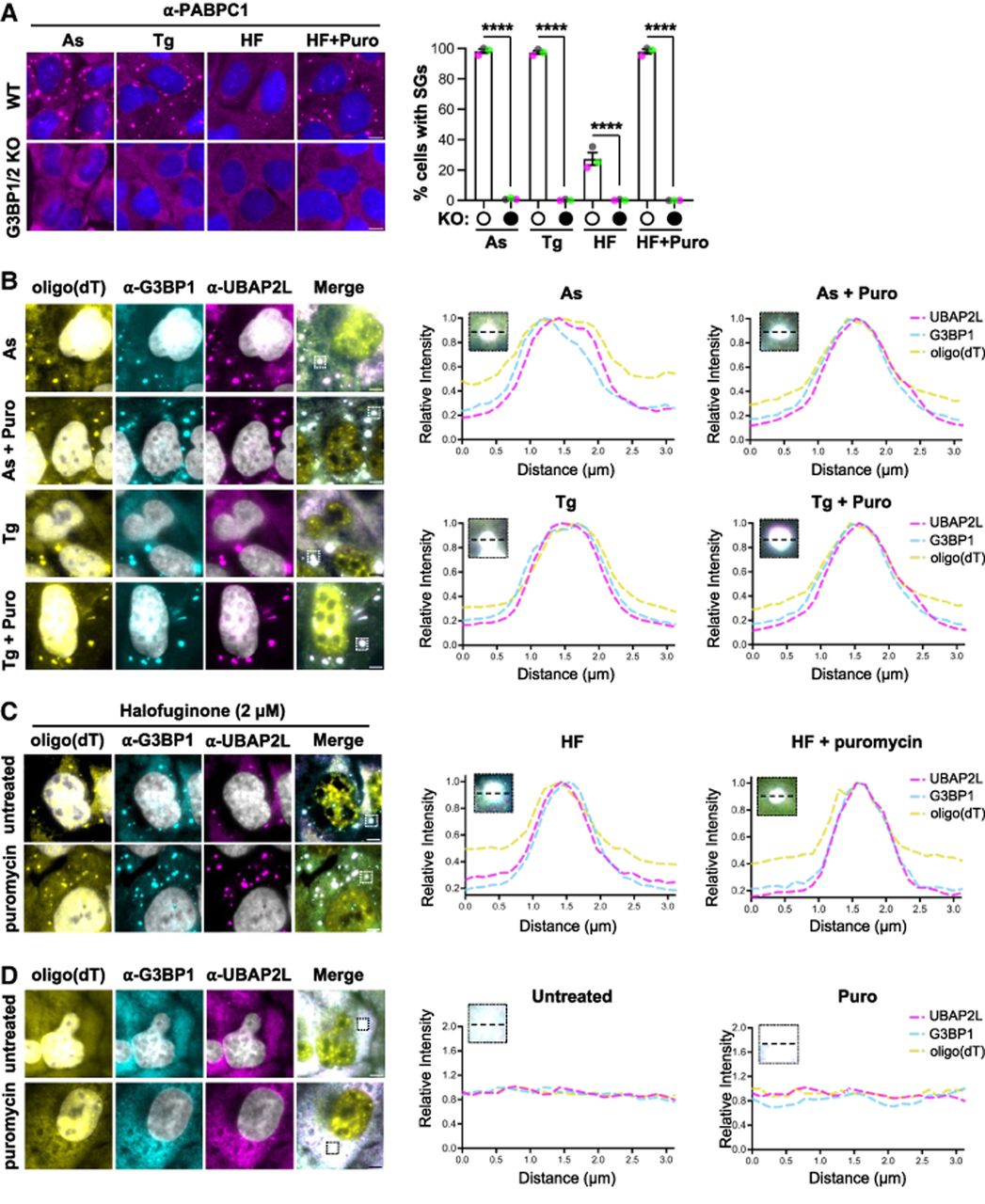
Halofuginone-induced stress granules require G3BP1/2 and contain canonical stress granule markers. (A) Stress granules (PABPC1 immunofluorescence; magenta) in wild-type or G3BP1/2 knockout U-2 OS cells treated with arsenite (250 µM, “As,” 1 h), thapsigargin (1 µM, “Tg,” 1 h), or halofuginone (2 µM, “HF,” 4 h) with or without puromycin (10 µg/mL) for the last 30 min. Nuclei stained with Hoechst (blue). Average percentage of cells with stress granules ± SEM with green, gray, and pink points representing the value from each replicate shown at right. Significance assessed with ordinary one-way ANOVA followed by Tukey's multiple comparisons test with (****) P < 0.001. (B) Representative images (left) of stress granule markers [polyadenylated RNA detected with oligo(dT) FISH, or G3BP1 or UBAP2L by immunofluorescence] in U-2 OS cells treated with arsenite (25 µM) or thapsigargin (0.04 µM) with or without puromycin (10 µg/mL). G3BP1 in cyan, UBAP2L in magenta, poly(A)+ RNA in yellow, and nuclei stained with Hoechst (gray). At right: Colocalization of stress granule markers (plot profile traces; peak for each channel set to 1) in a representative stress granule for each condition. (C) Stress granule markers in U-2 OS cells treated with halofuginone (2 µM, 4 h) with or without puromycin (10 µg/mL) during the last 30 min as in B. (D) Localization of poly(A)+ RNA, G3BP1, or UBAP2L as in B and C in unstressed U-2 OS cells in the presence or absence of puromycin (10 µg/mL, 1 h). All results represent n = 3 independent replicates. Scale bars, 5 µm.




