


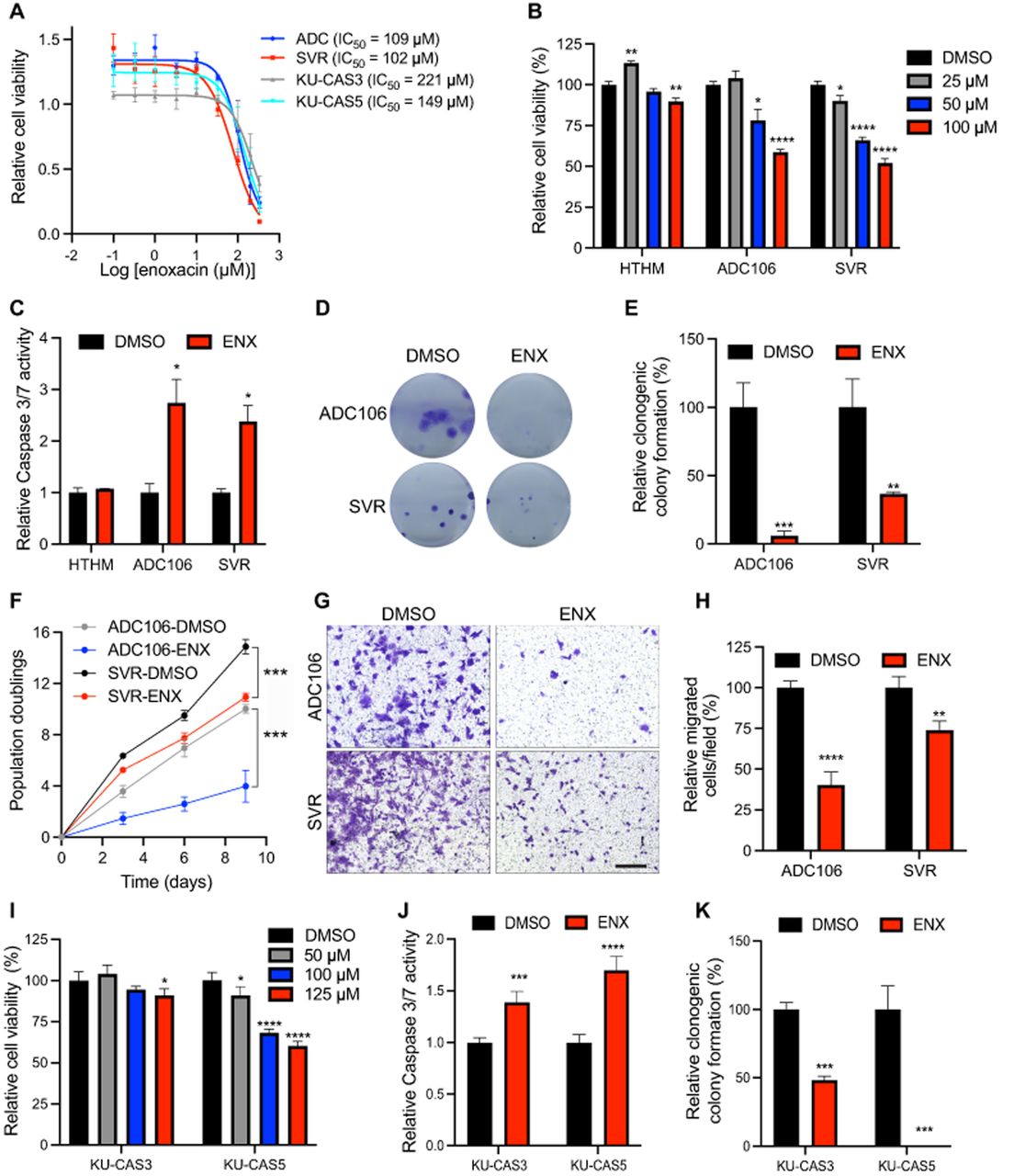
Enoxacin reduces angiosarcoma proliferation. (A) Cell viability curve based on CellTiter-Glo analysis of ADC106, SVR, KU-CAS3, and KU-CAS5 cells, 72 h after treatment with indicated concentrations of enoxacin (ENX). (B) Relative cell viability via CellTiter-Glo in HTHM, ADC106, and SVR cells treated with the indicated dose of ENX for 72 h. (C) Relative Caspase 3/7 activity in ADC106, SVR, and HTHM cells treated with DMSO or 100 µM ENX for 48 h. (D) Representative images and (E) quantification of clonogenic colony formation in cells treated with DMSO or 100 µM ENX. (F) Population doubling assays in ADC106 and SVR cells treated with DMSO or 100 µM ENX. (G) Representative image and (H) quantification of migrated cells in transwell migration assays in cells treated with DMSO or 100 µM ENX; scale bar, 150 µm. (I) Relative cell viability from CellTiter-Glo analysis of human AS cells treated with the indicated concentration of ENX for 72 h. (J) Relative Caspase 3/7 activity in cells treated with 125 µM ENX or DMSO for 48 h. (K) Quantification of clonogenic colony formation assays in cells treated with DMSO or 125 µM ENX, (*) P < 0.05, (**) P < 0.01, (***) P < 0.001, (****) P < 0.0001.




