


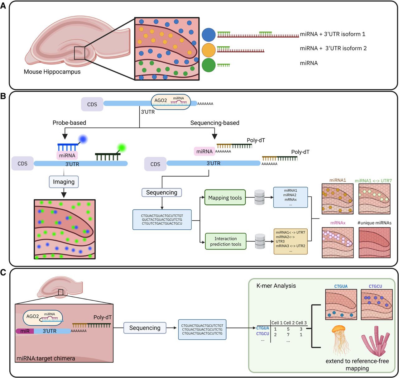
Methodologies for detecting the spatial context of miRNA-UTR interactions. (A) Conceptual illustration of miRNA abundance in the mouse hippocampus and interactions with different 3′UTR isoforms. (B) Overview of current state-of-the-art probe-based imaging (In situ hybridization) and sequencing-based (Poly-Adenylation) workflows. Sequencing-based methods rely on external databases (e.g., Human Genome, miRNA-target databases) and can identify spatial patterns of miRNAs, mRNAs and miRNA-3′UTR interactions in high-throughput. (C) Potential future workflows for miRNA-UTR spatial analysis include, on the experimental side, ligation-based approaches to construct miRNA:target chimeras directly on tissue capture arrays. On the computational side, k-mer analysis could be used to detect spatial patterns of sequences without relying on a reference genome, even enabling cross-species analysis.




