


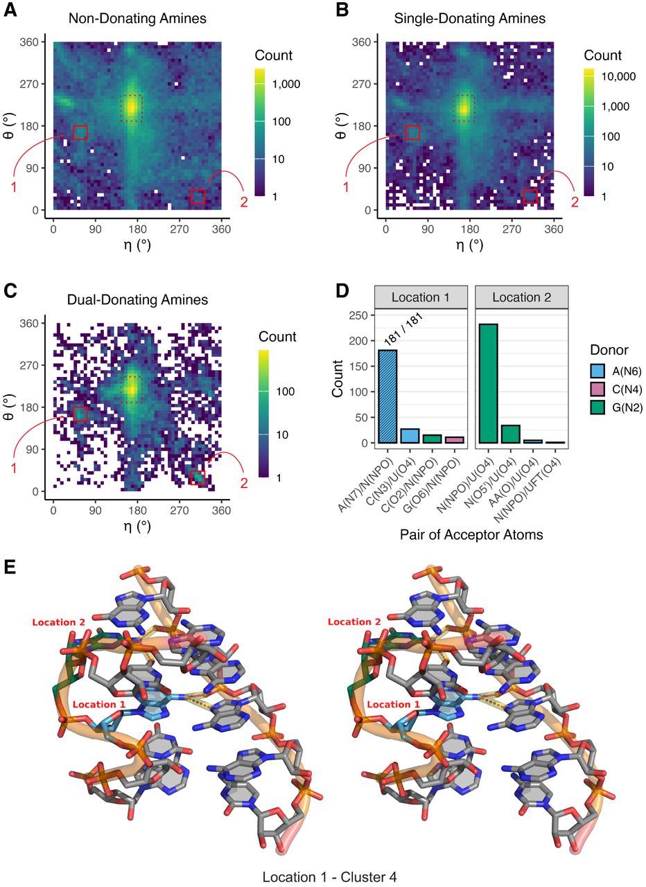
Key dual-donating amines involved in S-motifs. (A–C) Heat maps depicting pseudotorsion angles of residues bearing (A) non-, (B) single-, and (C) dual-donating amines. The color scale is logarithmic to accommodate the wide range of data. Regions that correspond to A-form helices are in dashed red boxes, and locations of interest, “Location 1” and “Location 2,” are in solid red boxes. The residues within Location 1 and Location 2 of (C) are provided within CSV and Python files; see Supplemental Table S1 and Supplemental Material. (D) Number of acceptor pairs that are engaged with dual-donating amines for Location 1 and Location 2. Symbols “N” and “AA” are used to denote any canonical RNA or amino acid residue, respectively, when the acceptor is a backbone atom. Only the top four acceptor pairs are shown for each Location. (E) Cross-eye stereoview of the representative structure from the dominant cluster (C4) (Supplemental Fig. S6) of the Location 1 analysis. An S-motif is depicted with an A dual-donating amine from Location 1 (blue; PDB ID 6ZU5, chain S60, A806) and a G dual-donating amine from Location 2 (green) (Ehrenbolger et al. 2020). Magenta dots highlighted in yellow depict H-bonds from the two dual-donating amines. All representative structures from all four clusters for Location 1 and Location 2 are depicted in Supplemental Figure S7.




