


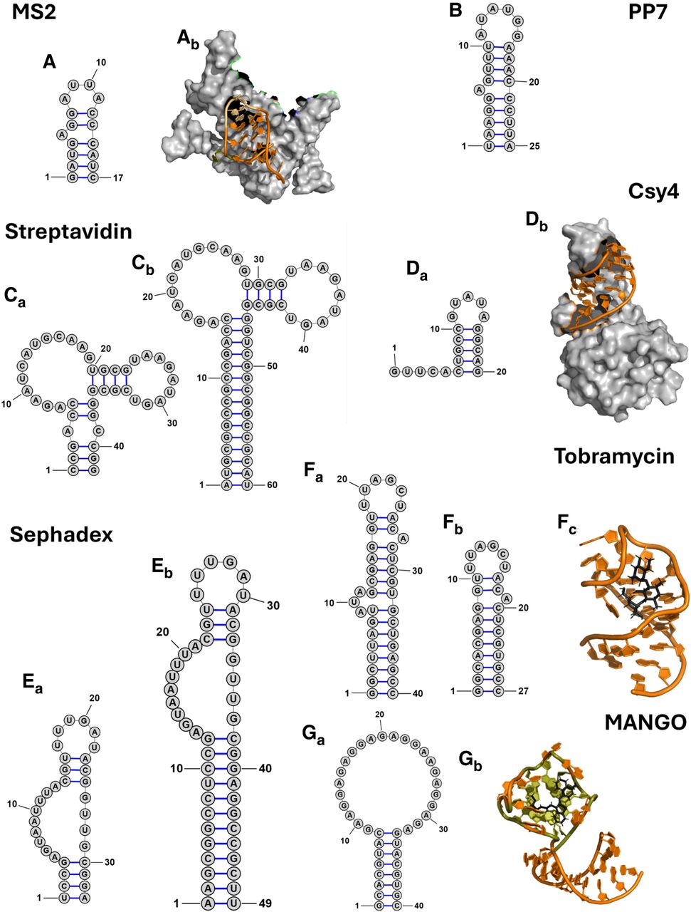
Comparison of aptamer structures used in RNP pulldowns. The secondary structures of aptamers are shown as predicted in the respective publications (Table 1). (A) (a) MS2 protein binding aptamer and (b) a 3D representation of the aptamer with the stem in orange, the bulged nucleotide in gold, and the loop region in yellow bound to the protein in gray (PDB ID 5MSF); (B) PP7 protein-binding aptamer; (C) variants of the S1 streptavidin binding aptamer, (a) S1 and (b) S1m; (D) (a) the Csy4 protein-binding hairpin, (b) a 3D representation of the aptamer (PDB ID 4AL5); (E) variants of the Sephadex binding aptamer (a) D8 and (b) D8m; (F) variants of the tobramycin-binding aptamer (a) j6f1, (b) J6, and (c) a 3D representation of the aptamer in orange bound to tobramycin in black (PDB ID 2TOB); (G) (a) the MANGOII aptamer and (b) a 3D structure of the aptamer in orange showing the G-quadruplex in gold and the ligand (thiazole orange-biotin) in black (PDB ID 6C65). Three-dimensional structures were visualized in The PyMOL Molecular Graphics System (Version 2.7, Schrödinger, LLC).




