


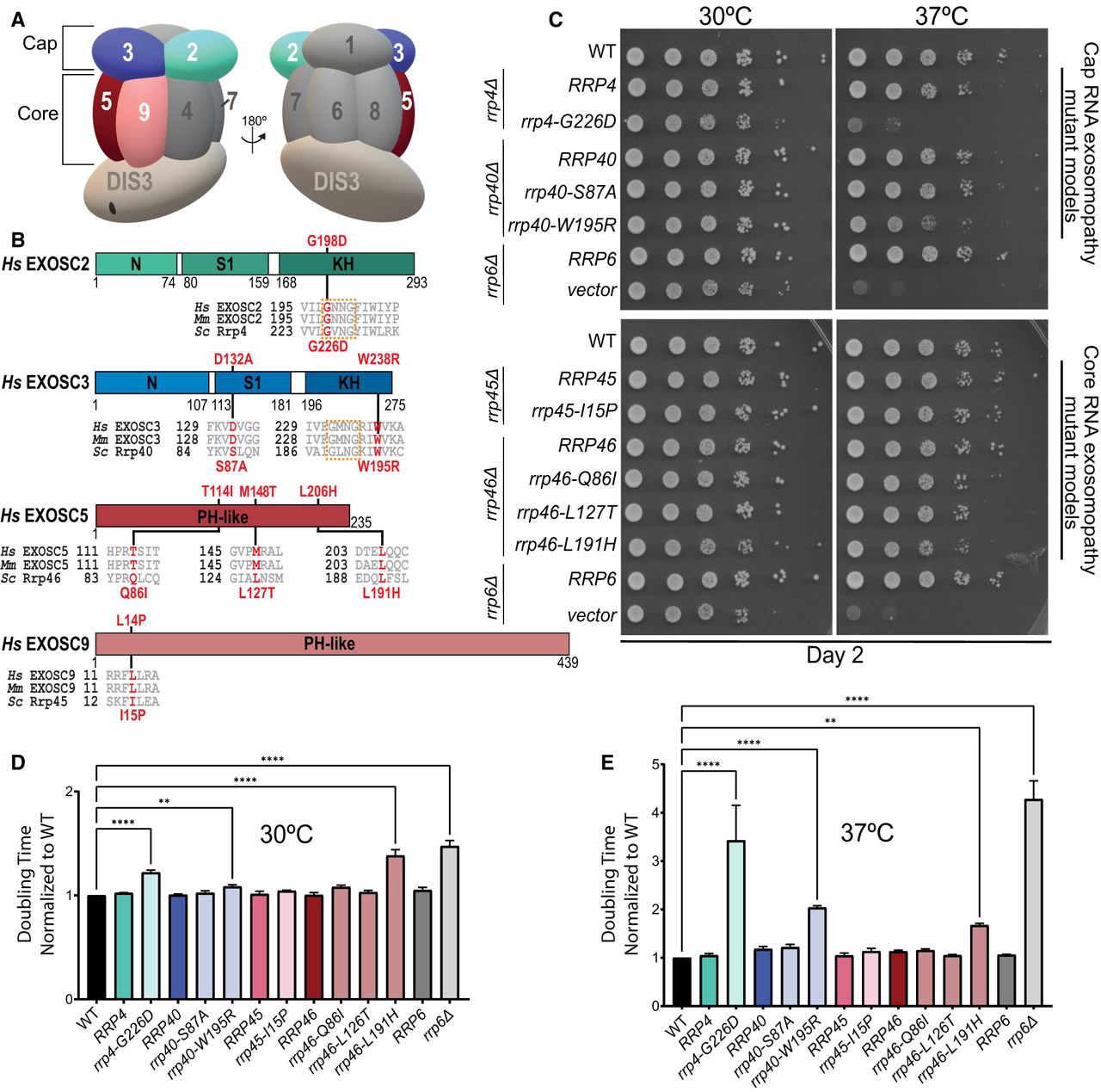
Overview of pathogenic amino acid substitutions in the human structural subunits of the RNA exosome. (A) Schematic view of the human RNA exosome with nine structural subunits (EXOSC1–9), denoted as 1–9, and one catalytic subunit (DIS3). (B) Domain maps of EXOSC2, EXOSC3, EXOSC5, and EXOSC9. EXOSC2 and EXOSC3 are composed of three domains: an N-terminal domain, a central putative RNA-binding S1 domain, and a C-terminal putative RNA-binding K homology (KH) domain. The “GxNG” motif in the KH domain of cap subunits is boxed in orange. EXOSC5 and EXOSC9 are composed of a singular PH-like domain. The positions of the RNA exosomopathy disease-linked amino acid substitutions in the human subunits are depicted above the domain structures in red. Sequence alignments of the orthologs from Homo sapiens (Hs), Mus musculus (Mm), and S. cerevisiae (Sc) reveal the high degree of conservation of the residues altered in disease (in red) and the sequences flanking these residues (in gray). The amino acid substitutions generated in the budding yeast Rrp orthologs for this study that correspond to the pathogenic amino acid substitutions are shown below the sequence alignments in red. The rrp4-G226D, rrp40-W195R, and rrp46-L191H mutant yeast cells, modeling EXOSC2-G198D, EXOSC3-W238R, and EXOSC5-L206H variants, respectively, exhibit (C) impaired growth in a solid media assay and (D,E) increased doubling times calculated from a liquid media assay at (D) 30°C and (E) 37°C. For these assays, the growth of RNA exosome deletion strains solely containing wild-type or mutant RNA exosome plasmid was analyzed by serial dilution and spotting of cells onto SD -Leu media and in liquid media, at 30°C and 37°C. BY4741 cells were used as wild-type isogenic controls (labeled WT). The rrp6Δ deletion containing an empty vector or wild-type RRP6 plasmid was used as a control. Growth measurements in liquid media used three biological replicates.




