


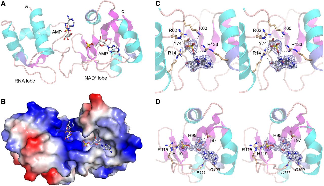
Structure of PhoTpt1 in complex with 5′-AMP. (A) Tertiary structure of PhoTpt1 (cartoon model) with two bound AMP nucleotides (stick models with gray carbons and yellow phosphorus atoms). One AMP is bound to the RNA lobe, and one AMP occupies the position of the NAD+ adenylate in the NAD+ lobe. (B) Surface electrostatic model of PhoTpt1 bound to the two AMP nucleotides (stick models). (C) Stereo view of the AMP in the RNA lobe overlaid with a simulated annealing omit density map (blue mesh contoured at 1 σ), highlighting Tpt1 hydrogen bond contacts to the 5′-PO4 and 3′-OH (black dashed lines). (D) Stereo view of the AMP in the NAD+ lobe overlaid with a simulated annealing omit density map (blue mesh contoured at 1.5 σ), highlighting Tpt1 hydrogen bond contacts to the adenine nucleobase, ribose hydroxyls, and 5′-PO4.




