


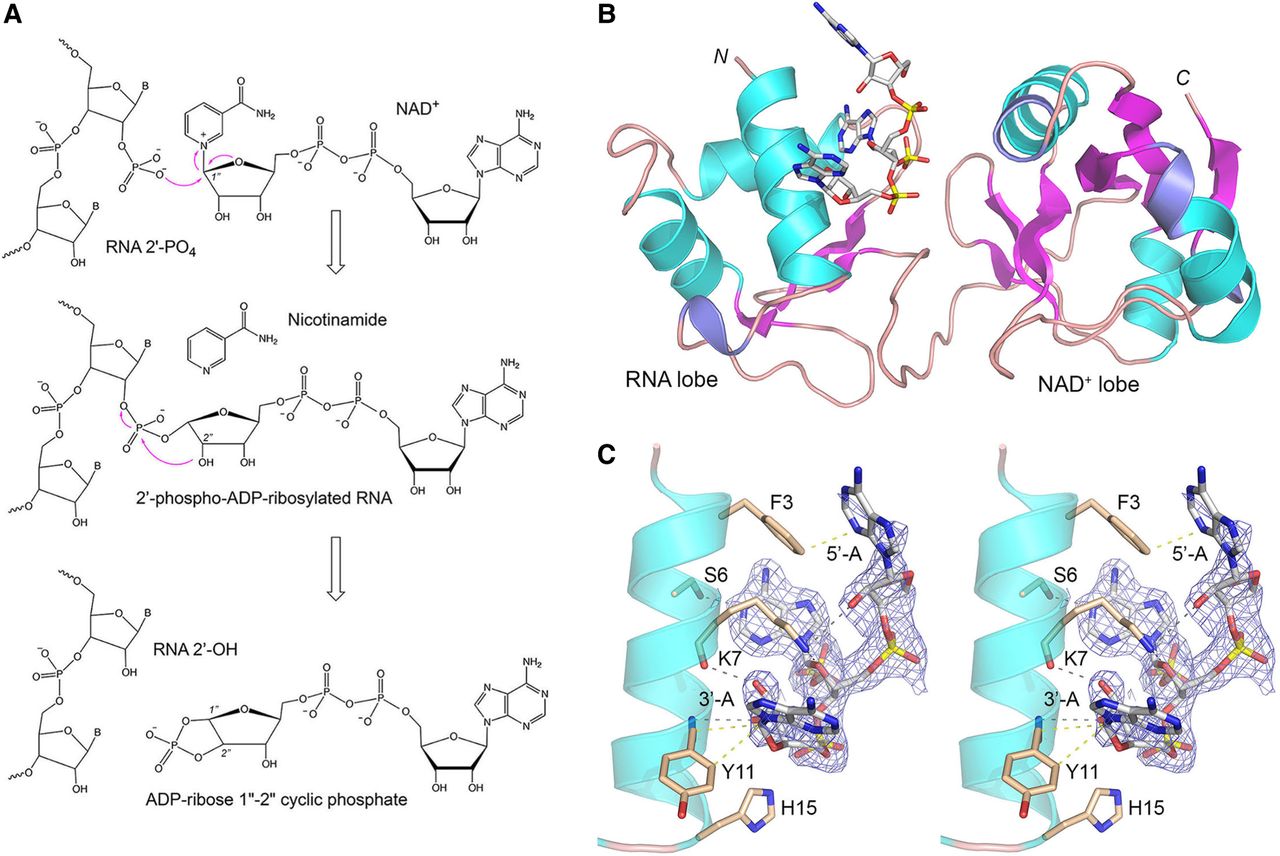
Tpt1 reaction mechanism and structure of PhoTpt1 in complex with 2′-PO4 RNA. (A) The Tpt1 reaction pathway comprises the two chemical steps shown in which: (i) the RNA 2′-PO4 reacts with NAD+ to expel nicotinamide and form a 2′-phospho-ADP-ribosylated RNA intermediate; and (ii) transesterification of the ADP-ribose 2″-OH to the RNA 2′-PO4 displaces the RNA 2′-OH and generates ADP-ribose-1″,2″-cyclic phosphate. (B) Tertiary structure of PhoTpt1, depicted as a cartoon model with magenta β-strands, cyan α-helices, and blue 310 helices. The ApA2′PpA substrate (stick model with gray carbons and yellow phosphorus atoms) occupies the RNA lobe. (C) Stereo view of the ApA2′PpA substrate overlaid with a simulated annealing omit density map (blue mesh contoured at 1 σ), highlighting its atomic interactions with amino acids in the N-terminal α1 helix of PhoTpt1 (depicted as stick models with beige carbons). Hydrogen bonds are denoted by black dashed lines. Van der Waals contacts are depicted as beige dashed lines.




