


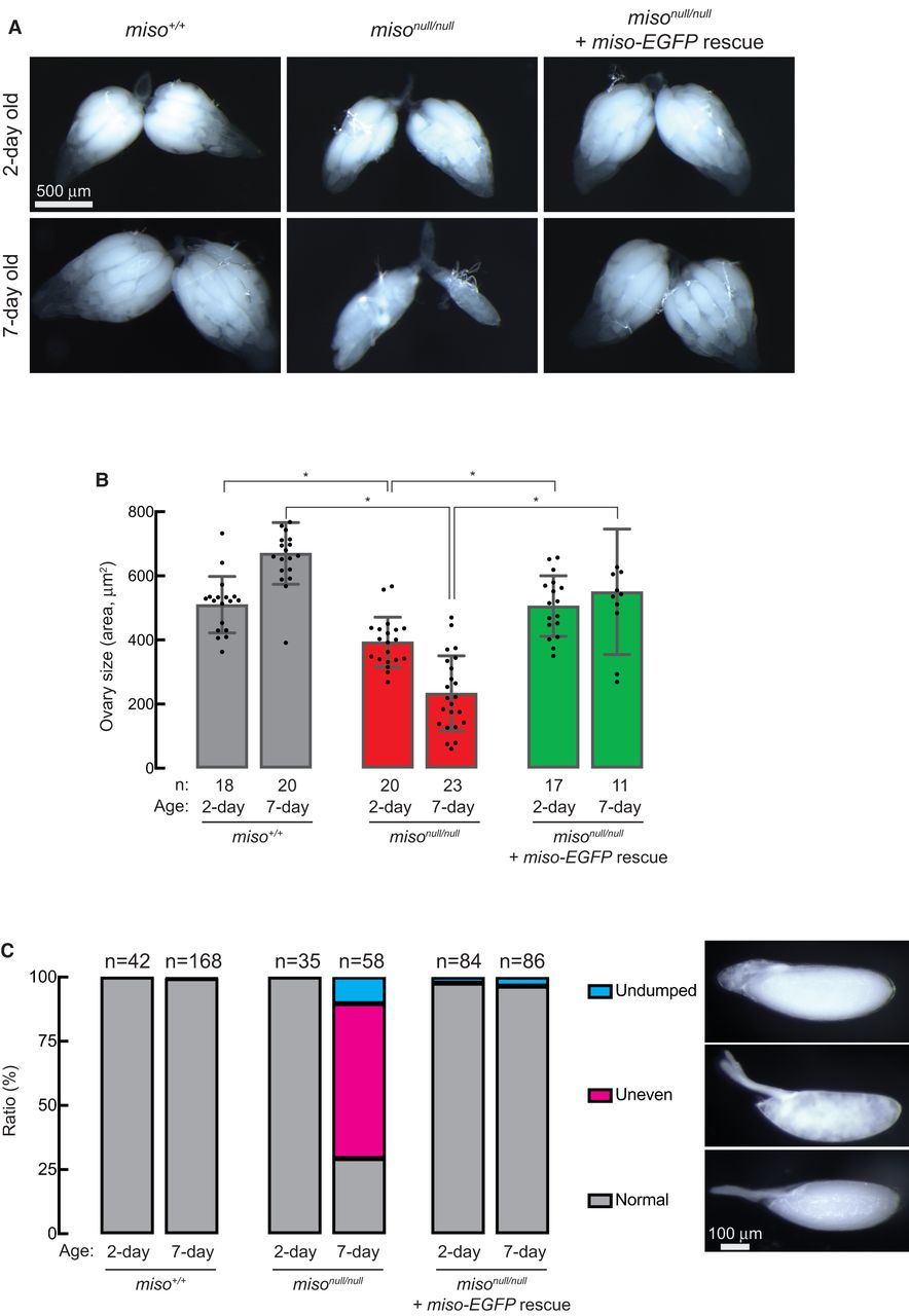
FIGURE 3.
Reduced ovary size and abnormal stage 14 oocytes in miso mutant females. (A) Stereomicroscope images of dissected ovaries. Scale bar, 500 μm. (B) Quantification of ovary size (area) from dissected samples. Mean ± SD. P-value < 0.05 (Student's t-test, unpaired, two-tailed) is indicated by *. (C) Percentage of stage 14 (mature) oocytes with normal, uneven, and undumped phenotypes. Representative images of these phenotypes are displayed. Scale bar, 100 μm.




